
Introduction
Malaria is the most important of parasitic diseases of humans and remains today, as it has been for centuries a large burden on tropical communities. Cerebral malaria is the most important complication of falciparum malaria and also the leading cause of death in malaria 1. It is defined as an acute, symmetric encephalopathy associated with sequestration of parasite-infected erythrocytes in the cerebral vessels and capillaries in patients with falciparum malaria 2.
WHO Criteria for Diagnosis
The World Health Organization has laid down definite guidelines for diagnosis and management of cerebral malaria 3. This definition requires the presence of unarousable coma using the Glasgow Coma Scale (GCS), exclusion of other encephalitides, especially bacterial meningitis and if possible, locally prevalent encephalitis and the presence of asexual forms ofP. falciparumin the blood film.
Retinal Involvement in Cerebral Malaria
Eye being an extension of the brain, retina provides a unique opportunity to observe the central nervous system vasculature and to study cerebral vascular pathology directly thus providing a clue to the mysteries of cerebral malaria. The detection of malarial retinopathy can be a candidate diagnostic test for cerebral malaria (Figs. 1-3). There is a set of retinal abnormalities that is unique to cerebral malaria. These abnormalities include blurred disc margins, papilloedema, retinal hemorrhages, retinal whitening, retinal edema, vascular changes and soft exudates. Of these retinal whitening and vascular changes are specific to cerebral malaria.
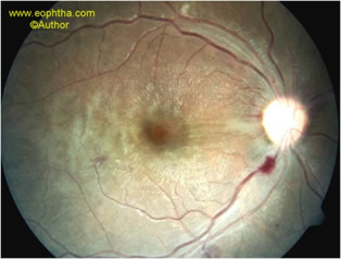
Fig. 1. Color fundus photograph of an adult, recovering from cerebral malaria, shows retinal whitening, retinal hemorrhage along the inferotemporal retinal vessel, cotton-wool spot adjacent to the superotemporal vessel and an isolated hemorrhage temporal to the macula.
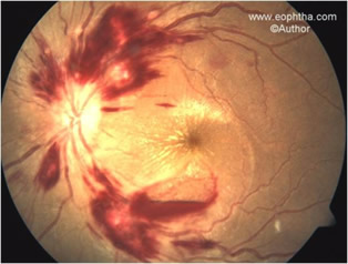
Fig. 2. Color fundus photograph of an adult, recovering from cerebral malaria, shows blurred disc margins, vascular occlusion in the form of retinal hemorrhages along the retinal vessels, cotton-wool spots adjacent to the superotemporal vessel, macular fan and an isolated preretinal hemorrhage inferior to the macula.
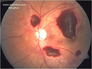
Fig. 3. Color fundus photograph of an adult, recovering from cerebral malaria, shows multiple preretinal hemorrhages as well as superficial retinal hemorrhages.
Retinal whitening:Retinal whitening referred to as retinal edema in earlier reports, can be macular whitening or peripheral whitening. Macular whitening is a patchy opacification of the retina centered on the fovea, sparing the central fovea, or foveola and frequently extending temporally between the vascular arcades. Peripheral whitening is similar in appearance, does not cross blood vessels and occurs in a more mosaic pattern in the peripheral fundus. The appearance is distinct from cotton wool spots, which also occur, but less frequently. Retinal whitening is similar in appearance to patchy ischemic retinal whitening, an uncommon finding in central retinal vein occlusion, but has a different retinal distribution.
Vessel changes:An orange or white discoloration of retinal vessels is found in peripheral fundus, involving either discrete sections of vessels, or peripheral trees. Larger vessels exhibit white or orange tramlining which can be continuous or interrupted, delineating an apparently narrowed blood column. A widely distributed whitening of retinal capillaries and post-capillary venules may occur, making them prominent against the choroidal background. Though associated with a fatal outcome these are not independent predictors of fatal outcome in cerebral malaria.
Hemorrhages:Retinal hemorrhages in malaria are predominantly white-centered, intra-retinal, blot hemorrhages similar to Roth spots. In severe cases, these can be extremely numerous (>120 in each eye) and overlapping. Flame shaped, blot and boat shaped hemorrhages of varying sizes also occur frequently. Occasionally, hemorrhages can extend into the pre-retinal space. However neither the number nor the evolution of hemorrhages is an independent predictor of worse outcome.
Papilledema:Papilledema is not specific to malaria and can occur in many other conditions that cause brain edema and coma. It accompanies retinal features of cerebral malaria in a proportion of cases and independently increases the risk of fatal outcome. When papilledema is present without retinal whitening, vessels changes, or white-centered hemorrhages, the examiner should consider other causes of raised intracranial pressure.
Cotton wool spots:Cotton wool spots in malaria though occur less frequently, are better demarcated, more brightly white, and less widely distributed than retinal whitening.
Hard exudates:Hard exudates are rarely found in malarial retinopathy.
Retinal edema:Retinal edema in malaria is found to be associated with coexistent disc hyperemia and blurred disc margins.
Blurred disc margins:These may be associated with retinal hemorrhages, disc hyperemia or pallor and retinal edema.
Pathogenesis
The characteristic histopathologic feature ofP. falciparummalaria is sequestration of parasitized erythrocytes in microvessels by cytoadherence. This cytoadherence is due to the appearance of high molecular weight adhesive proteins on erythrocytes’ surface causing them to stick to the receptors on venular and capillary endothelium. Apart from this infected RBCs adhere to uninfected RBCs to form rosettes and to parasitized RBCs (agglutination). The processes of cytoadherence, rosetting and agglutination play a central role in the pathogenesis. It has been seen that eyes that had vessel whitening in life had sequestration in retinal vessels at autopsy, with cytoadherent erythrocytes containing late stages ofP. falciparumand little hemoglobin 4. It is only the blood column of retinal vessels that is normally seen at ophthalmoscopy, and erythrocyte hemoglobin is the pigment responsible for their red color. It is believed that sequestered erythrocytes whose hemoglobin has been consumed by parasites account for tramlining, and for the orange and white appearance of vessels.
The number of retinal hemorrhages seen on fundoscopic examination correlates with the number of cerebral hemorrhages in fatal cerebral malaria 5. In common with cerebral hemorrhages, fibrin thrombi are seen in the small vessel at the center of hemorrhages and hemorrhaged red blood cells rarely contain parasites.
These suggest that macular whitening is caused by oncotic swelling of second-order neurones in the inner retina due to metabolic or hypoxic stress. A study using fundus fluorescein angiography suggests that metabolic steal by intravascular parasites may play a role, rather than capillary obstruction, which was not seen in association with macular whitening. The typical distribution of macular whitening, in sites of high metabolic demand and in vascular watershed zones, supports causation by metabolic steal or hypoxia over toxic malarial products, cytokines, or nitric oxide.
References
- WHO practical chemotherapy of malaria. Technical report 805. Geneva, World Health organization, 1990, p 7.
- Macpherson GG, Warrell MJ, While NJ, Looareesuwa S, Warrell DA. Human cerebral malaria: A quantitative ultrastructural analysis of parasitized erythrocyte sequestration. Am J Path. 1985; 119: 385-410.
- Warrell DA, Molyneux ME, Beales PF. Severe and complicated malaria. Tran R Soc Trop Med Hyg. 1990; 84 (Suppl 2): 1-65.
- Lewallen S, White VA, Whitten RO, Gardiner J, Hoar B, Lindley J, Lochhead J, McCormick A, Wade K, Tembo M, Mwenechanyana J, Molyneux ME, Taylor TE. Clinical-histopathological correlation of the abnormal retinal vessels in cerebral malaria. Arch Ophthalmol 2000;118: 924–928.
- White VA, Lewallen S, Beare N, Kayira K, Carr RA, Taylor TE. Correlation of retinal haemorrhages with brain haemorrhages in children dying of cerebral malaria in Malawi. Trans R Soc Trop Med Hyg 2001;95: 618–621.