
Diplopia charts are kept in postgraduate examinations during viva sessions. Even in theory examinations also, these topics may be asked. It is not difficult to interpret the charts and all PGs should practice recording the results of these charts.
Let us see the method of interpretations of these charts
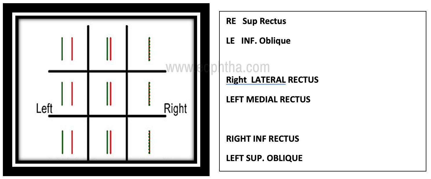
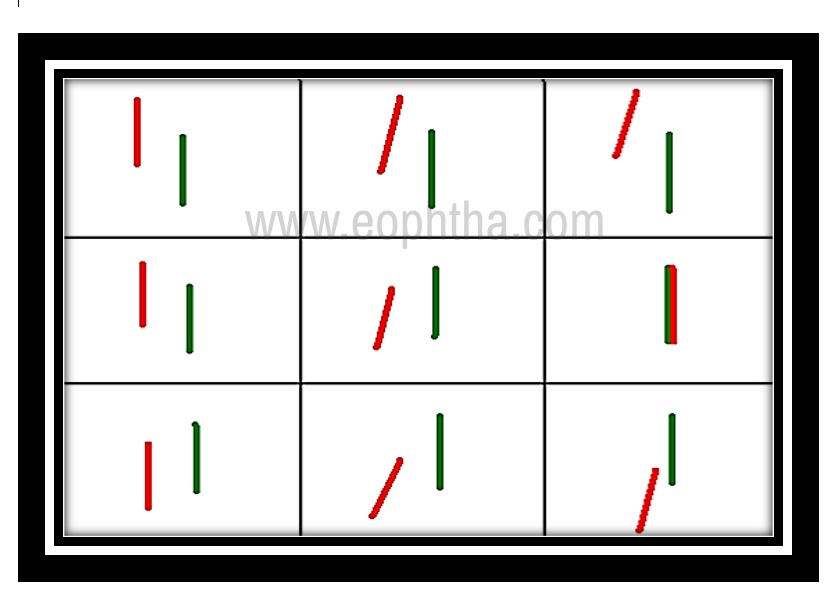
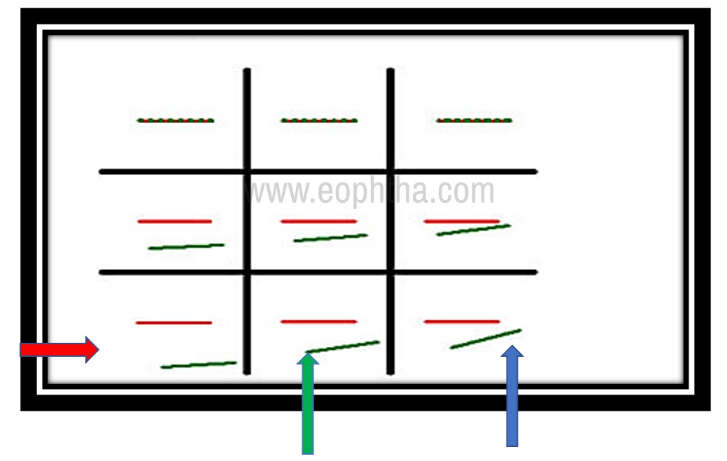
- See the chart
- Remember red and green goggles are used. Red in front of the right eye and Green in front of the left eye.
- To your right side (The right side of the picture). Version positions.
- Dextrum version. Elevation and Depressions are there.
- The left side of you is the left side positions like levoversion, elevation and levodepression are there
- Primary position elevation and depressions are noted.
- Totally we have nine quadrants
- Never forget to write the name of the muscles of the right eye and the left eye working in the various positions. For example in dextroelevation, the right eye superior rectus and left eye inferior oblique must be written as shown in the above chart.
As in the below chart write the muscles in levoposition
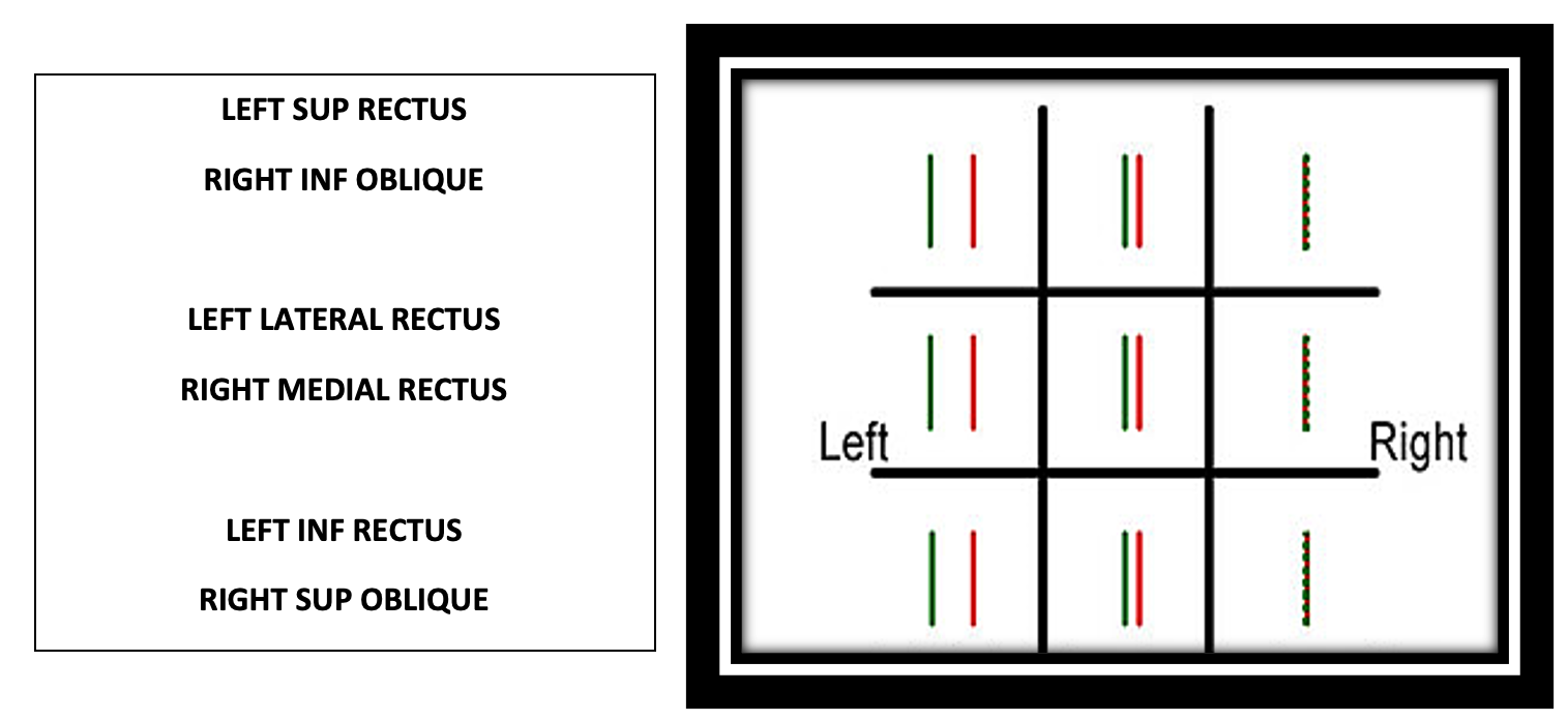
First never forget to remember the yolk muscles working in the particular quadrant
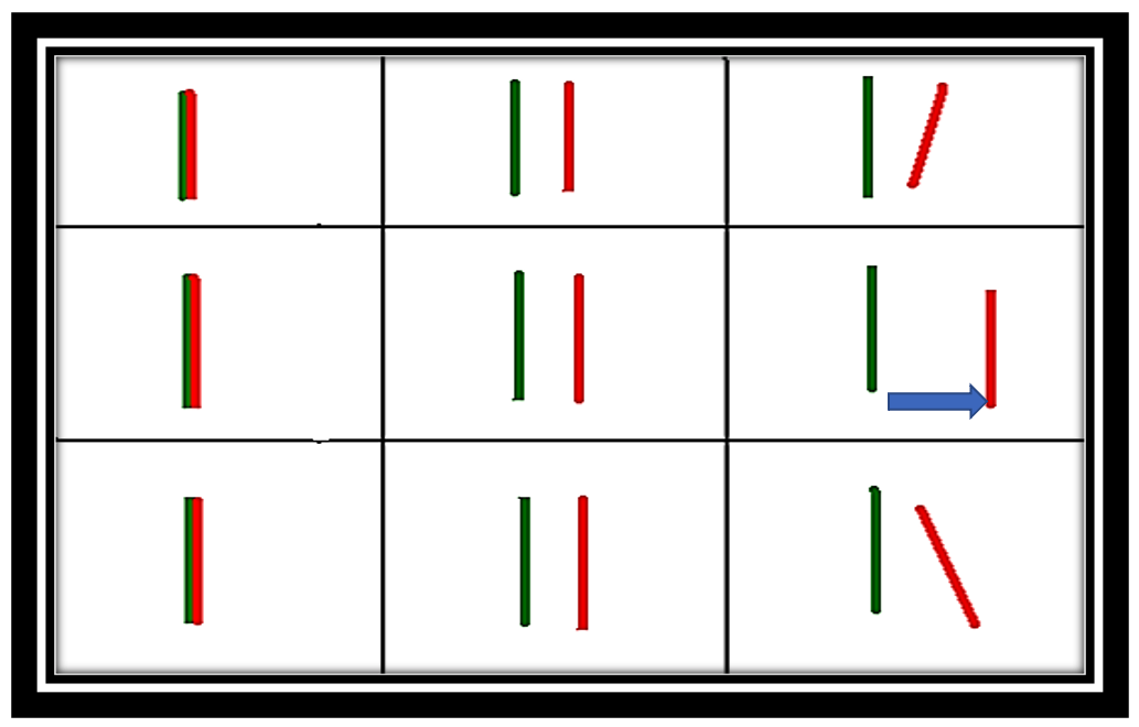
Find out which color image is away from the center of the chart, in other words, which image is present outside in the version quadrants or in vertical positions
For example, in the above chart, a green image is present outside in the levopositions
That means the left eye has a problem
See for the horizontal or vertical separations. Here only horizontal separation only- that means only horizontally moving muscles are affected
Notice Green image is present on the left side or right side (uncrossed or crossed)
Here green is to the left side only. Uncrossed diplopia.
In dextropositions, no diplopia
Uncrossed diplopia because of problem in horizontally moving muscle of the left eye. There are only two muscles like medial rectus and the lateral rectus.
Always remember when abductors are paralyzed, the patient will experience uncrossed diplopia (Abductors: Lateral rectus, Superior oblique, Inferior oblique)
In this picture only horizontal muscle is involved because of horizontal position diplopia is there. Uncrossed means, lateral rectus of the left eye is involved
IF MEDIAL RECTUS MEANS CROSSED DIPLOPIA WILL BE SEEN
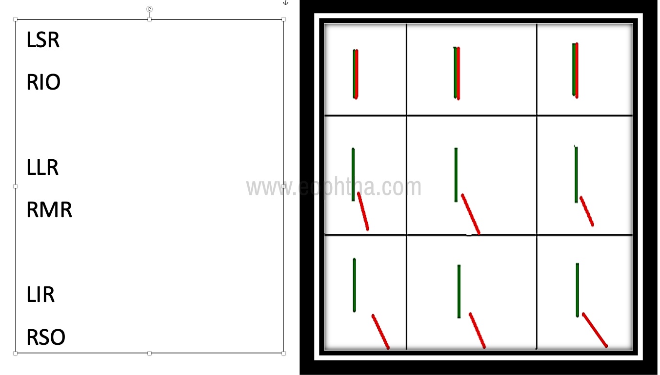
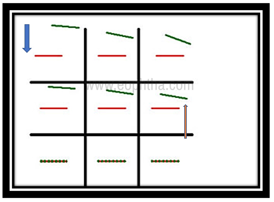
We have discussed the problem related to horizontally acting muscles. Let’s look into the vertically acting muscles with subsidiary horizontal as well as torsional movements.
The name of the yolk muscles should be written on the side of the chart.
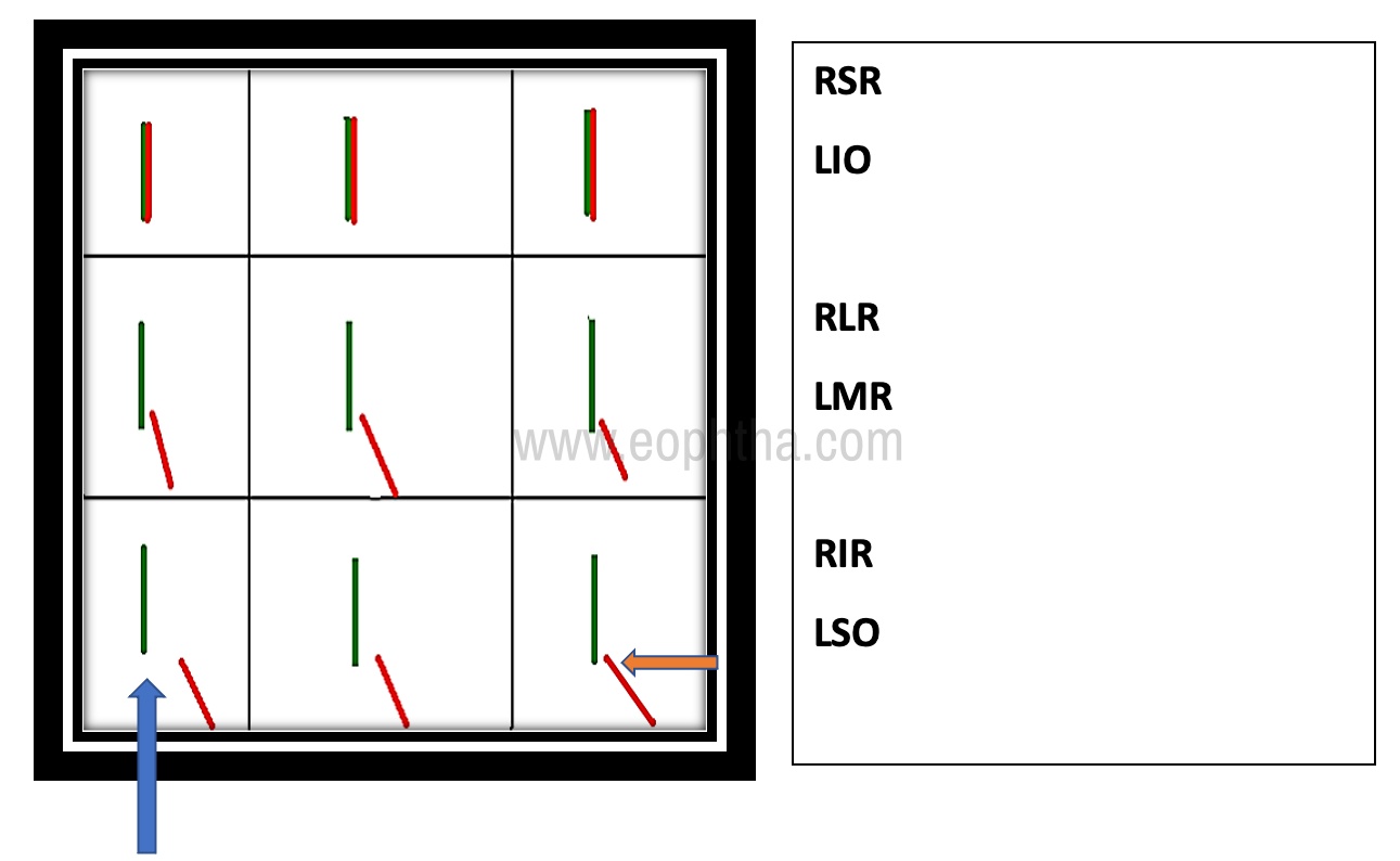
In this above chart, see which color image is away from the center of the chart. Here the red is downwards which means the depressor of the right eye is affected. (Don’t forget that red glass in front of the right eye). Next check the image to find out whether it is crossed or uncrossed. Here the red image is towards the right side only. So uncrossed. Right eye depressor uncrossed is superior oblique.
See the maximum separation in levodepression. Positions where the superior oblique of the right eye is taking part in the movement ( BlueArrow)
The other depressor in the right eye inferior rectus will produce crossed diplopia. See the tilt of the image. Here it is (Orange Arrow) leaning to the left side
Always remember the tilt will be towards the sound side ( normal side ) when the superior named muscles - superior oblique and superior rectus ( Intortors ) are paralyzed.
So, the diagnosis here is right eye superior oblique paralysis
Let us explore some more examples:
Look at the picture below: red is away from the center. So, the problem is in the right eye. All positions crossed except in dextroversion where the lateral rectus is working: Right 3rd nerve palsy
Red in an upper position elevated - Elevator palsies
Red in depressed in downwards positions - Depressor palsies
Horizontal separation is also seen
Red is present on the left side of the chart. It is crossed diplopia.
Crossed diplopia can occur in adductors problems. Medial rectus, Superior and Inferior recti. Never forget Recti. ( SR, IR) are adductors).
Simply remember the third nerve problem in this chart. Here in the third nerve problem, the tilt of the image is always to the affected side only
No diplopia in dextroversion where the lateral rectus of the right eye is taking part. Here diplopia is present everywhere except in the dextroversion position
Remember
6th Nerve Anomalies: Uncrossed Diplopia
4th Nerve Anomalies: Uncrossed Diplopia
3rd Nerve Anomalies: Crossed Diplopia
Red image away from the center. So, the right eye is having problem,
Only horizontal separation and the red image is on the right side - uncrossed,
The right lateral rectus is involved. Uncrossed diplopia is produced.
Maximum separation in dextroversion (Blue Arrow)
Don’t bother here about the tilts shown
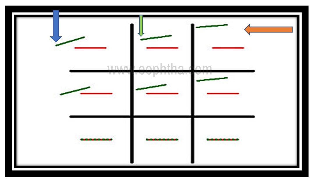
Here green away from the center. So, left eye problem.
Elevated. Elevators in the left eye are involved.
Green slightly going towards to right side crossed (OrangeArrow)
Maximum separation levoelevation (Blue Arrow) See here the image tilting towards the sound right side (Orange Arrow)
In Superior rectus palsy, the tilt of the image is to the sound side
Diagnosis: Left Superior Rectus Palsy
Look here, the green image away, so the left eye involved
- Elevated. Elevator problem
- Green coming towards the left. Uncrossed (Blue Arrow)
- Maximum separation in dextroelevation area (Orange Arrow) only Elevator uncrossed is inferior oblique.
- Tilted to affected side ( left side) ( Ipsilateral side) (Green Arrow)
Remember
Inferiors named muscles if paralyzed (inferior rectus & inferior oblique): Tilt to Ipsilateral side
Superiors named muscles if paralyzed (superior rectus & superior oblique): Sound side tilt
Green away, so left eye problem
Depressed: Depressor of the left eye is involved
Green coming towards the right. So, crossed (Blue Arrow)
Maximum separation in Levodepression ( Red Arrow)
See the image is tilted to the left side ( Ipsilateral side) ( Green Arrow)
Diagnosis: Left inferior rectus palsy

Green away from the center, so, left eye is involved
Depressed so, depressor of the left eye is involved
Green coming towards the left. So, uncrossed (Blue Arrow)
Maximum separation in dextrodepression (Green Arrow)
Only uncrossed depressor is superior oblique in Left eye.
See here the tilt towards the sound side ( Right side) (Orange Arrow)
Diagnosis: Left superior oblique palsy