“If you are brave enough to start the right way, you will be strong enough to finish at the right place.”
The most crucial aspect of vitreoretinal surgery is learning it the right way. And if you have the guidance of teachers with experiences of a lifetime, there is an abundant pool of knowledge you can extract. Plenty of young VR surgeons are facing difficulties in the basic principles and doctrines of surgery. Some of these basic questions were compiled together and addressed to the top-notch VR surgeons of India, owing to a simple and lucid explanation for the same.
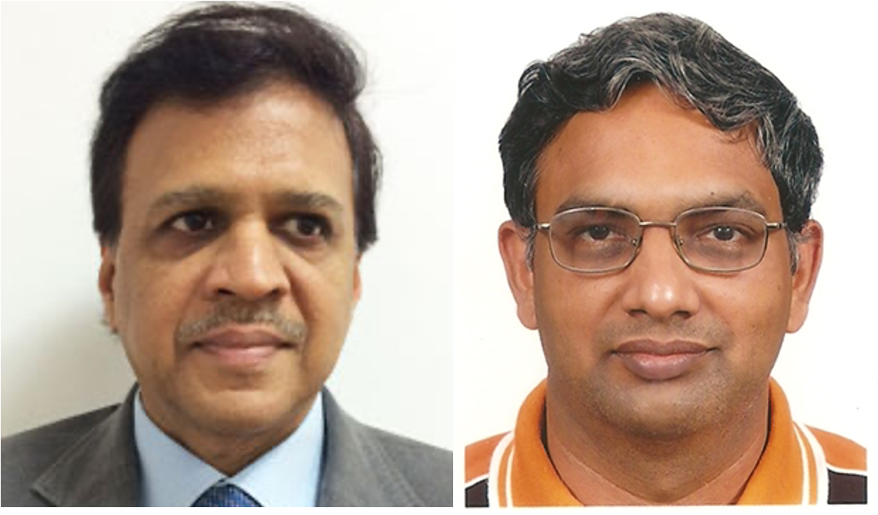
Dr. Atul Kumar, MD, FAMS, FRCS(Ed) is the Chief & Professor of Ophthalmology at Dr. R.P. Centre, AIIMS, New Delhi since 1st January 2016. He completed MD & Sr. Residency from Dr. R.P. Centre, AIIMS New Delhi India & joined as Assistant Professor in the same institute in 1987 (Discipline: -Vitreous-Retina). He pursued Retina Fellowship from the University of Maryland, Baltimore, USA, 1990. He has 313 indexed & non-indexed publications and has edited a book on “RETINA: Medical and Surgical Management” 2018, 1st edition, Jaypee Bros Publishers. He has chapters in over 24 books. He has been awarded “Padma Shri” in 2007 for contribution to healthcare in the field of Vitreo-Retinal Diseases and Surgery by the President of India. He is Appointed Hony. Advisor Ophthalmology to Govt. of India. Since May 2016. He was also awarded FRCS Edinburgh by Royal College of Surgeons 2017.
Dr. Pramod S Bhende, MS, is currently the Director ofShri Bhagawan Mahavir Vitreoretinal services at Sankara Nethralaya, Chennai, India. He has delivered over 370 presentations in various national and international conferences and published 82 papers in peer-reviewed national and international journals. He is a recipient of 4 named orations. His special interests include surgery for retinopathy of prematurity, diabetic retinopathy, and combined procedures involving anterior and posterior segments pathologies. He is a member of the National Task Force for ROP. He is actively involved in teaching and training VR fellows at Sankara Nethralaya.
Here we have an exclusive interview, with Dr. Atul Kumar and Dr. Pramod Bhende, who provide us answers to some of the basic fundamentals of vitreoretinal surgery.
eOphtha: What is your take on ergonomics in VR surgery? What special measures do you personally take in order to maintain a fine spinal balance?
Dr. Atul Kumar: Ergonomics is very important while performing a VR surgery. Vitreoretinal specialists have to work in repetitive, non-ergonomic postures that stress the musculoskeletal system.A recent survey of retina specialists showed that 85% of respondents had affected neck or back pain. First of all, one should always take care of getting the patient’s position and table height adjusted. The head of the patient should be comfortably positioned at the end of the table, to avoid the need for leaning forward. We use a well-designed VR chair which allows the surgeon to adjust height with an electronic foot pedal and also has adjustable armrests. Lastly, the microscope should be adjusted with the alignment of the angle of oculars at the surgeon's eye level, to avoid neck flexion or extension. The surgeon should assume a neutral spine position with normal curvature, throughout the procedure. I personally find ‘Ngenuity’ (by Alcon Inc.) 3D heads up viewing system for VR viewing very much more ergonomically sound. The surgeon takes a heads-up position which allows a straight back and neck.
Dr. Pramod Bhende: Unlike most anterior segment surgeries, VR surgeries are of longer duration and proper posture during surgery is essential to avoid back and neck problems and to prolong professional carrier as a VR surgeon. To achieve the best possible outcome, it is important that the surgeon should be sitting comfortably in his/her chair and relaxed while operating. The correct height of the operating table, comfortable chair with arm-rests and adjustable height is very much needed. The height of the chair should be adjusted in such a way that the arms with 90 degrees bent at elbow should be at the level of the patient’s eyes/ forehead. The elbows should be resting/ supported on chair arms and the wrists should be resting on the draped forehead of the patient. The back and neck of the surgeon should be straight /erect. The legs should be bent at 90 degrees and comfortably resting on-foot controls. All the foot controls should be placed on the ground in such a way that the surgeon should be able to access them without stretching his/her legs. In an institutional setup, where more than one surgeon is using the same operation room settings, the surgeon can use additional supports, if needed, to maintain the desired posture. The microscope should be in a neutral position before focusing at the pupillary plane of the patient. Adjusting inter-pupillary distance for microscope eye-pieces is also equally important. With the surgeon maintaining the primary gaze, the microscope eyepieces should be adjusted at the level of the surgeon’s eyes. To avoid fatigue and hand tremors, the grip on the instruments should be firm but not very tight. Regular back and neck stretching exercises are also equally important to maintain back muscles’ tone.
PEARL: Make sure you and your patient are completely relaxed and comfortable. The height of the chair should be adjusted in such a way that the arms with 90 degrees bent at elbow should be at the level of patient’s forehead. Avoid neck flexion or extension. Adjust the eyepiece, IPD, and foot control. The grip on the instruments should be firm but not very tight. And most importantly, stretch and exercise from time to time.
eOphtha: Some surgeons prefer to do a diabetic vitrectomy with a 25/27G instrument, while a retinal detachment with a 23G instrument owing to the ease of air-fluid exchange. Do you decide on the gauge of the instrumentation depending on the type of case you are dealing with, or are you gauge-specific for all surgeries?
Dr. Atul Kumar: I usually decide the gauge of the instrumentation depending on the type of case I am dealing with. Specifically, for a complicated diabetic TRD, I prefer to do bimanual vitrectomy with chandelier’s light, and I prefer a combination of 23G and 27G vitrectomy with a cut-rate of 10,000 cuts/min. This allows correct identification of surgical plane and bimanual dissection of firmly adherent fibrovascular membranes and thick, taut hyaloid without damaging the retina. A 27-gauge vitrectomy cutter allows working very close to the retina while avoiding iatrogenic breaks, while 23 G vitrectomy helps to perform the peripheral dissection.
Dr. Pramod Bhende: With better fluidics and technology, using any of the gauges is OK. Surgeons’ comfort and the availability of the instruments is an important factor in decision making.
But in certain situations, the selection of a specific gauge is important. In highly myopic eyes, 23G is preferred due to a longer axial length of the eye. Similarly, if you are planning to do lensectomy for hard nuclear cataract, Framatome is available only with 20 or 23 G system. Cases where surgeons anticipate excess manipulation, anterior or extreme peripheral dissection, 27G should preferably be avoided as instruments are more flexible. For membrane peeling 25 or 27 G is better, especially 10K beveled probe, for ease of getting the correct plane for dissection.
PEARL: Decide the gauge of the instrumentation depending on the type of case you are dealing with. It’s imperative to be flexible with all gauges.
eOphtha: What are the important measures you take while adjusting the BIOM and the patient’s eye in order to obtain the optimal view during the surgery?
Dr. Atul Kumar: Non-contact optical systems such as BIOM require adjustments to ensure good visualization for safe and effective surgery. Securing the patient's head in a head ring and positioning it parallel to the floor should be the first step. The microscope should be centered using the reset button before focusing on the microscope. Take care while lowering the condensing lens to avoid hitting the cornea. After putting viscoelastic over the cornea, lower the condensing lens as close as possible to the cornea. I do not use these non-contact viewing systems routinely in my surgeries and prefer the Mini-Quad XL lens by VOLK® which allows an extended field till over 130 degrees and panoramic view and find it a very surgeon –friendly contact WAVS.
Dr. Pramod Bhende: At present, there are multiple wide-angle visualization systems available in the market. Grossly they can be divided as contact and non-contact systems. The most commonly used non-contact systems are BIOM and Resight. For focusing BIOM, I take the following steps:
- Correct assembly of BIOM and especially alignment of the condensing lens is to be checked
- The microscope should have a position at a central / neutral position.
- Meticulous sticking of the drape around the surgical area to ensure that air does not leak from the drape edges. This will avoid the fogging of the condensing lens of BIOM during surgery.
- Instead of irrigating solutions, use dispersive viscoelastic to coat the cornea during surgery to prevent drying. This will prevent fluid splashing on the condensing lens.
- Once BIOM is in place, activate stereo diagonal inverter (SDI).
- Use fine focusing screw to get sharp image of the surgical field. The surgeon has to keep refocusing intermittently to maintain crisp focus of desired surgical field as surgery progresses. With newer models of BIOM, footswitch can be used to adjust focus during surgery.
- Position condensing lens as close to the cornea as possible to get the largest possible surgical field.
- When BIOM is in position, focusing mechanism of the microscope is used to change the surgical field (increase or decrease).
- Zoom function of the microscope will continue to get magnified image of the surgical field.
PEARL: Understand and get familiarized with focusing mechanism of your visualization system before starting surgery. Make sure your microscope is at neutral first. Clean and align your viewing system accordingly. Use viscoelastic for coating cornea. Most importantly, if you don’t see, don’t blindly cut!
eOphtha: Do you feel that the modern-day retinal detachment surgery is shifting from buckles to vitrectomy? Do you feel it’s an industry drive revolution?
Dr. Atul Kumar: Vitrectomy has greatly evolved over the years and has significant advantages. With the advent of small gauge 23/25G micro-invasive vitreoretinal surgery (MIVS), vitrectomy has become a lot safer and can ensure complete removal of vitreous. For the surgeon, it allows clear visualization of retinal pathology like retinal breaks, especially suited for posterior breaks, large or multiple breaks, myopic retinal detachment (RD) with the thin sclera, and is a faster procedure. For the patient undergoing vitrectomy, postoperative pain is much less with faster recovery, lesser dry eye, less post-operative astigmatism, and superior cosmesis. However, the role of encirclage with vitrectomy is still relevant in RD with severe proliferative vitreoretinopathy (PVR) changes, especially in the inferior quadrant Even in primary phakic eyes, base-shaving seems easily possible. I do not think this is industry-driven.
Dr. Pramod Bhende: Well, partly yes to both! Scleral buckling has a long track record with a more than 95% success rate. With improved technology, we have newer instruments making vitrectomy procedure more safe and predictable with better anatomical and functional outcome. MIVS also helps in early wound healing due to smaller incision size hence reduced hospitalization and early mobilization of these patients. Various studies have shown that both the techniques are equally good, though there are specific groups where one technique would be better than the other. For example, vitrectomy scores over scleral buckle (SB) in pseudophakic eyes but SB has a better success rate in phakic eyes with good media clarity. I personally feel that the selection of the surgical procedure should be case dependent. But the choice should be left with the operating surgeon. He/ she is the best person to decide which technique would be better in his /her hands to get the best possible outcome in a given case.
PEARL: The recovery period with a newer MIVS system is definitely better. Having said that, various studies have shown that both the techniques are equally good for retinal attachment, though there are specific groups where one technique would be better than the other. Thus, the selection of surgical procedures should be case dependent.
eOphtha: What tips would you give to prevent a lens touch while performing laser to the superior retina in phakic patients?
Dr. Atul Kumar: Positioning the superonasal and supero-temporal ports closer to the 3 and 9 o’clock positions allow a good range of movement and better access to superior retina. The endolaser probe is curved and allows performing laser to superior retina quite easily. The probe should not cross to the other side just behind the central portion of the lens as it is thickest. It can lead to lens touch with the shaft of the laser probe. Prefer doing laser using the same side port. Peripheral indentation can be done to do laser of anterior retinal areas.
Dr. Pramod Bhende: Best way is to use curved or angled laser probe to laser superior peripheral retina. Lens touch is not really an issue while lasering superior retina between two sclerotomies. The real problem is visualization and reaching to the superior retina. Visualization also gets compromised in a meridian of sclerotomy with endo-illumination due to inadequate light reaching the retina immediately surrounding the cannula. By rotating the eye in superonasal and/or supero-temporally to some extent one can manage to complete the laser. In eyes with preexisting buckle, it will be difficult to visualize and reach retina on the posterior slope of the buckle. Angled laser probe is the best in this situation. While working with MIVS and trocar cannula system, instruments can be exchanged in any of the sclerotomies to easily approach desired part of the retina.
PEARL: Keep the upper two sclerotomies 150-170 degrees apart. Used curved or angled laser probes for periphery. Avoid crossing the midline with the probe in phakic patients to avoid lens touch. Rotation of the eyeball and peripheral indentation are some tricks to do superior laser.
eOphtha: A lot of young surgeons today tend to use PFCL in all cases of retinal detachment, be it simple or complex. What is your take on that aspect?
Dr. Atul Kumar: PFCL is very useful while operating on GRTs, RD with severe PVR, posteriorly dislocated lens, RD with macular holes, ILM autografts. PFCL acts as a second hand of a VR surgeon. Membrane peeling becomes much more safe and stable. In macula on RDs, photoreceptor displacement is also reduced at the macula. However, it should be used judiciously in selected cases to avoid possible disadvantages of retained subfoveal PFCL and to make surgery cost-effective.
Dr. Pramod Bhende: VR practice has significantly changed after PFCL was introduced. Indications for PFCL usage are evolving every day. It’s indeed a great surgical tool and made intraoperative maneuvering much simpler in a select group of VR disorders. However, I personally feel that it is not necessary to use PFCL for every case. A majority of the cases can be managed without it.
PEARL: PFCL is an aid, not the solution. It has indeed has made intraoperative maneuvering much simpler in a specific group of cases. However, adverse effects like subretinal migration, incomplete removal, and photoreceptor toxicity should be kept in mind. Thus, judicious use, in select cases, is the key.
eOphtha: After the fluid-air exchange, the view tends to get very hazy and it gets difficult performing the endolaser. Sometimes the air enters the anterior chamber, or bleeder ooze causes view blockage. Any tips or tricks to tackle such scenarios?
Dr. Atul Kumar: Preoperative evaluation can be very helpful. The detailed anterior segment should be done to assess any posterior capsule defect (larger than size of optic of IOL), weakened/ broken zonules, and stability of IOL. In these cases, OVD like healon can be injected in anterior chamber to avoid air getting into anterior chamber. For bleeders, bleeding points should be carefully diathermized before starting fluid air exchange, at low infusion pressure like 10 mm Hg. Sudden massive bleeding can also be controlled by raising intraocular pressure to 60 mm Hg. Air will itself act as a tamponading agent at high pressures. Sometimes, fluid has to be again started and bleeders should be coagulated and FAX has to be repeated again.
Dr. Pramod Bhende: My tips are:
- Hazy view after FGE in a phakic eye can be due to the drying effect of air on the posterior capsule of crystalline lens. This can happen when the surgeon is waiting for the laser machine to get connected after FGE. To avoid waiting, try and keep the laser ready before starting FGE and complete the laser as quickly as possible after FGE(before lens become hazy).
- In a pseudophakic eye, the view can get compromised due to condensation on the posterior IOL surface, especially when there is a defect in posterior capsule. Coating the posterior IOL surface with viscoelastic will help to improve the visualization.
- In an aphakic eye, compromised visualization can be due progressive miosis due to prolonged pupillary contact with air and also due to Descemet folds. Coating corneal endothelium will improve the visualization of the vitreous cavity. Also, finish the laser as quickly as possible before pupil constricts.
- In a phakic or pseudophakic eye, if air enters in the anterior chamber or if there is bleeding, it can be replaced by filling the chamber with viscoelastic to complete the posterior segment maneuvering. The viscoelastic can either be left behind or can be washed out at end of the surgery depending on case to case. Constrict the pupil at the end of surgery to minimize the possibility of reentry of air or silicone oil in the anterior chamber in post-operative period.
PEARL: Preoperative evaluation of lens/IOL status, zonules, dilatation, media clarity etc. is extremely crucial. Make sure your laser is working well before starting the FGE procedure. Use of OVDs in the anterior chamber can help if there is a recurrent entry of air. If there are bleeders, either cauterize at low IOP, or raise the bottle height. Avoid operating in a vague field as far as possible.
eOphtha: There is a constant emphasis these days on early removal of silicone oil in primary non-complex retinal detachment surgeries. What is your take on that?
Dr. Atul Kumar: Silicone oil can be toxic to the retina with complications like secondary glaucoma, cataract and band-shaped keratopathy, in case it comes in the AC. Once oil gets emulsified, it is usually difficult to remove the entire oil. So, early silicone oil removal (approximately 3 months) is advisable in primary non complex retinal detachments after ruling out PVR changes and before oil emulsification sets in.
Dr. Pramod Bhende: Silicone oil is used in an eye as an internal tamponade. After retinopexy, firm chorioretinal adhesions are formed around retinal break/s generally between 10 days to two weeks. Theoretically, silicone oil removal can be planned any time after that if the retina is well settled and intra ocular pressure is within normal range. However inflammation and proliferative process takes much longer time to stabilize and varies depending on etiology of the detachment. Also there are certain logistic, psychological and financial issues one needs to address while planning early second surgery. Hence I personally would delay SOR at least for 3 to 4 months after primary surgery unless there is a compelling reason for early removal of silicone oil.
PEARL: Early silicone oil removal at approximately 3-4 months is advisable in primary non-complex retinal detachments after ruling out PVR changes and before oil emulsification sets in.
eOphtha: A lot of young surgeons today talk about perfecting cataract surgery first and then entering a VR training program. What is your take on that?
Dr. Atul Kumar: Retinal surgery requires perseverance, strength, precision and dexterity. There is a steep learning curve. There is a lot of gentle tissue handling and precise micro-movements. Cataract surgery can give residents a chance to master on tissue handling, surgical manipulations and manual dexterity. So, it is advantageous if a person is good at cataract surgery. However, it is not a hard and fast rule that one has to attain excellence in cataract surgery to start vitreoretinal training. One needs to be proficient in tissue handling and precise hand movements.
Dr. Pramod Bhende: Combined cataract surgery and a vitrectomy procedure is always a challenging situation. V R surgeon can always invite one of his anterior segment colleagues to help to take care of cataract. Although not absolutely necessary, having good training in both anterior and posterior segment is always beneficial. Equipped with this knowledge, it is easier for the surgeon to decide about the best possible surgical plan in a given case and eventually helping the patient to get the best possible outcome.
PEARL: It is definitely advantageous if a person is thorough with cataract surgery before entering VR training, as it can aid in deciding an overall surgical plan and have proficiency in tissue handling, surgical manipulations, and dexterity. However, it is not hard and fast rule that one has to attain excellence in cataract surgery to start vitreoretinal training.
eOphtha: What tips would you give to the freshly passed out surgeon regarding case selection for VR surgery in his initial days of practice?
Dr. Atul Kumar: One needs to start operating simpler cases like Vitreous hemorrhage with PVD etc in the early part of the career to gain confidence. Learning proper wound construction is also paramount for starting MIVS. Patients should be adequately counseled before the surgery about prognosis and postoperative instructions should be emphasized for better patient compliance. Working under the supervision of senior trained surgeons can also help you evolve in your surgery and practice.
Dr. Pramod Bhende: Most important is to know your limitation and facilities /set up you have with you. A thorough evaluation of each case is very important. There is no scope for short cuts. To choose relatively technically simpler cases which as a surgeon one can handle confidently.
It is advisable that the surgeon himself/herself should do ultrasonography and other ophthalmic investigations, if needed, before taking the patients for surgery for a better understanding of patho-anatomy, surgical planning, and better intra-operative orientation.
Vitreous hemorrhage (and no underlying retinal detachment) with PVD or with single/isolated attachment, retinal detachment without PVR or cases with only posterior pole pathology and clear media are the typical cases that can be handled easily by newer surgeons. Once the surgeon becomes confident and gets used to his settings, more and more difficult/complicated cases can be taken for surgery.
PEARL: Simple cases in the early stages are the key to gain confidence and long term success. The setting you are working in also plays a major role. Understanding the tissue takes time. Meanwhile, master the art of counseling well. And always keep learning. Success is a ladder, not a jet-pack, do not rush in. And that’s the bottom-line!