
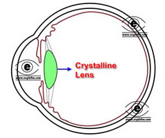
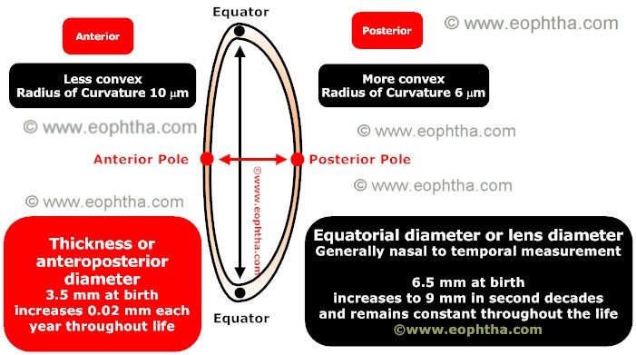
The crystalline lens is a unique transparent, biconvex, avascular intraocular structure with slightly more curved posterior surface .The radius of curvature of anterior surface is 10 mm and that of posterior surface is 6 mm.

Lens is a unique structure, which contains cells of a single type, in various stages of differentiation
Topography:
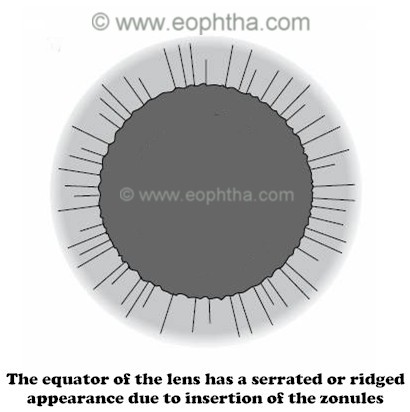
The center of the anterior surface is called anterior pole and it is situated 3mm away from the posterior (endothelial) surface of cornea. The center of the posterior surface is called posterior pole. The distance between these poles is measured as lens thickness. The thickness of lens is 3mm at birth, which increases to 6mm in older age. The marginal circumferences of the lens, where anterior and posterior surface meet, are known as equator. Equatorial or lens diameter is generally measured in nasal to temporal dimension. The equatorial diameter of lens is 6.5 mm at birth, which reaches to 9-10 mm in adult life. The equator of the lens is encircled by the cilliary processes of the cilliary body and held in position by zonules (see later) laterally. The distance between lens equator and cilliary processes is 0.5mm. Lens equator has a serrated or ridged appearance which is caused by the zonular fibers and this serration or ridges disappears during accommodation due to relaxation of the zonular fibers.
Refractive power:Location:
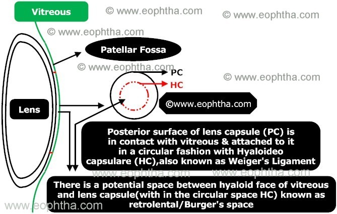
The Lens is situated behind the iris and in front of the vitreous. The posterior lens surface is attached to anterior vitreous in a circular fashion by Hyaloideo capsulare (HC) which is also known as Weiger's ligament. It is not a true ligament and strength of the attachment decreases with age. The potential space between hyaloid face of vitreous and lens capsule which lies within the circular space of hyaloideo capsulare is known as Burger's space or retrolental space.
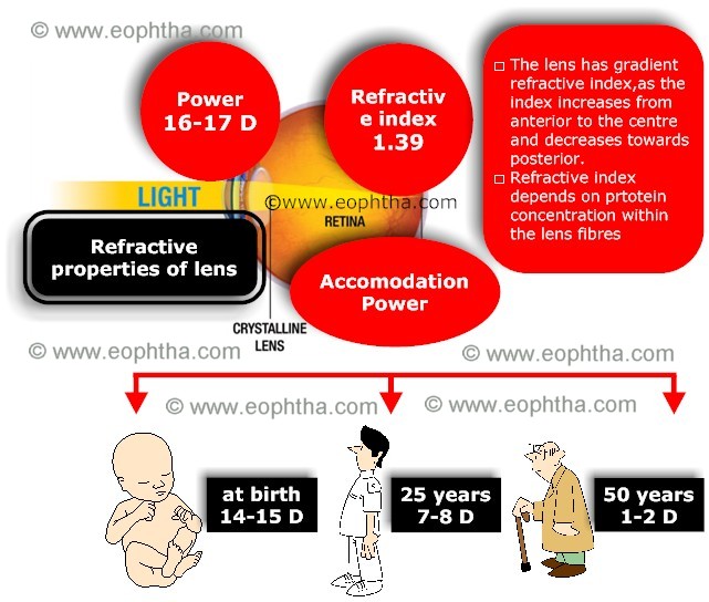
The diopteric power of human eye is approximately 58 diopters. The refractive power of crystalline lens is about 15 diopters. Though lens has less refractive power than cornea, it has the ability to change its shape with the help of cilliary muscle, by which it can change its diopteric power, allowing the distant and near vision. However this property changes with age. Lens has a refractive index of 1.39 (1.36 in periphery and 1.40 centrally - a property which is termed as grading refractive index)
Accomodative Power of Lens:
The eye has the capacityto adjust its focusfrom distance to near objects because of the ability of the lens to change shape, a phenomenon known as accommodation. The inherent elastic property of the lens allows it to become more or less spherical depending on the amount of tension exerted by the zonular fibers on the lens capsule. Zonular tension is controlled by the action of the parasympathetically innervated ciliary muscle
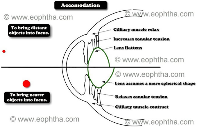
- When cilliary muscles contracts, relaxation of zonular tension occurs. The lens then assumes a more spherical shape, resulting in increased dioptric power which helps to bring nearer objects into focus.
- Ciliary muscle relaxation causes the zonular tension to increase. As a result, lens flattens, which helps in bringing more distant objects into view.

|
Structure of lens:
Lens is histologically composed of three structures-lens capsule, lens epithelium and lens fibers.
Lens Capsule:
Lens capsule is a transparent covering that surround the entire lens. Histologically it is a basement membrane, secreted by lens epithelium and lens fibers .The capsule is produced anteriorly by the lens epithelium and posteriorly by the elongating fiber cells. It is composed of type IV collagen fibers and sulphated glycosaminoglycans. Though it has no elastic tissue, it is highly elastic in nature because of lamellar or fibrillar arrangement of fibers. This property of the lens gradually decreases with age.
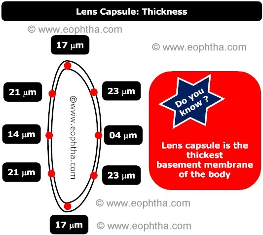
Lens capsule is thickest near equator and thinnest at posterior pole. Thickness of anterior lens capsule increases with age, whereas thickness of posterior capsule remains constant or changes slightly.
|
Capsulorhexis: Diameter of the adult lens is approximately 10mm and an area of diameter 6mm is the zonule free area in anterior capsule. In cataract surgery, a circular opening is made (capsulorhexis) with in this area. To overcome the elastic strength of the capsule, while performing continuous curvilinear capsulorhexis, two types of forces are applied-tearing by stretching (force is applied perpendicular to the desired direction of tear which is uncontrolled) and tearing by shearing (force is applied perpendicular to the capsular plane and it is more controlled).As the capsule in children is highly elastic than in adults, it becomes very difficult to perform continuous curvilinear capsulorhexis in such patients.
Lens epithelium:
The lens epithelium is a simple cuboidal epithelium and is found only in the anterior surface of the lens.Theses cells (A cells) secrete the anterior lens capsule throughout the life.
|
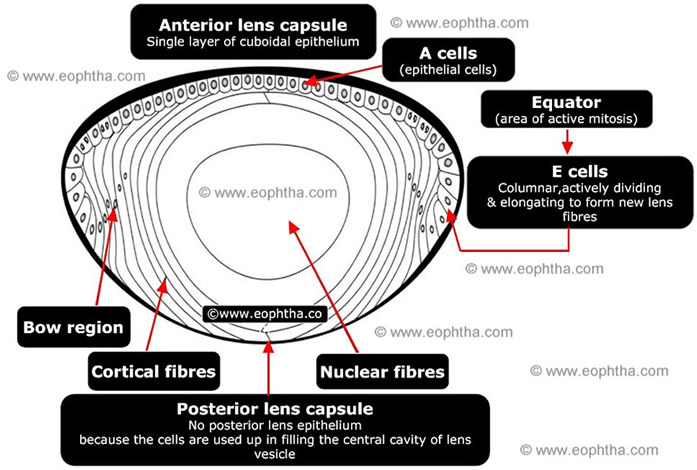
Near the equator.the cells of the anterior lens epithelium,elongate and becomes columnar(E cells).As they elongate their apical parts lies deeper to other cells which are placed more anteriorly.Thus these elongated epithelial cells trasformed to lens fibres at the equator. The band consisting of preequatorial and equatorial cells is called germinal zone.Here the mitotic capacity of the cells are at maximum. Cells in the germinal zone divides constantly.The newly formed cells are forced into the transitional zone where they elongate and differentiate to form the fiber mass of the lens.
There is no posterior lens epithelium because the cells originally located there have elongated into primary fibers of the lens
After cataract surgery, residual epithelial cells may cause posterior capsule opacification. E cells migrate posteriorly along the posterior capsule and often forms large balloon like bladder cells, known as Wedl cells.
Remnant cells on the anterior capsule after cataract surgery differentiate into spindle-shaped, fibroblast-likecells, which are known as myofibroblasts. They express smooth muscle actin filament, expressed commonly in smooth muscle cells and become highly contractile. These cells proliferate and migrate to the posterior capsule and form a layer by secreting extracellular ground substances and a basement membrane like material. Cellular contraction by this highly contractile cells leads to the formation of folds and wrinkles in the posterior capsule.
Lens Fibers:
As the transitional zone cells continue to elongate and differentiate, they turn meridionally. The apical end of these cells pass anteriorly towards the anterior pole and the basal end are pushed posteriorly towards the posterior pole. These processes of newly formed cells, pushing towards the center of the lens substance continues throughout the life. In this way with the growth of the lens, new superficial lens fibers are added in a concentrically arranged lamina, like the layers of an onion.
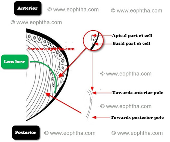
Now it is obvious that as the lens fibers elongate anteriorly, the nucleus of the cells also moves a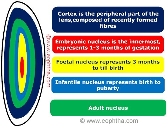 nteriorly. So the nuclei of the deeper cells will remain anterior to the nuclei of the more superficial cells. If a line (green line in picture) is drawn to connect nuclei of these cells, the line will take an arcuate shape anteriorly. These configurations of the nuclei are known as "lens bow". Gradually these cells loss all cellular organelles and becomes a lens fiber.
In cross section, each lens fiber is hexagonal in shape and may be up to 12 mm long. They are tightly packed together with the help of ball and socket type cytoplasmic interdigitations and gap junctions.
Zones of Lens:
Approximately nucleus occupies 84% of the lens and cortex occupies 16%.The nucleus is further subdivided into embryonic, fetal, infantile, and adult nuclei. Primary lens fiber cells, formed in the lens vesicle during embryogenesis forms the embryonic nucleus and the fibers laid down around the embryonic nucleus before birth forms the foetal nucleus. After birth, new fibers formed before puberty give rise to infantile nucleus and adult nucleus is formed after puberty. The cortex consists of recently formed nucleated fibers which lie outside the adult nucleus of the lens. The fibers of the cortex are loosely arranged whereas fibers of the nucleus are arranged in more compact fashion, the reason for which the nucleus is harder in consistency than cortex. Epinucleus is formed by the zone between foetal nucleus and cortex.
|
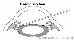
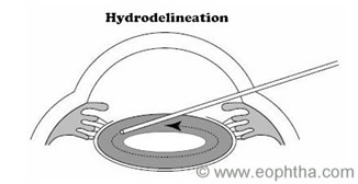
Hydroseparations
|
Sutures of the lens:
Junction of the lens fibres with other lens fibres of the same plane at the poles are known as suture of the lens.Thus anterior suture is formed by the apical parts of the lens fibres and posterior suture by the basal parts .During embryogenesis ,the lens fibres meet in three branches,there by forming a Y shape.The resultant anterior suture is an upright Y and the posterior one is an inverted Y.The sutures formed after birth has multiple branches like 6 to 9 or 9 to 15 and are of dendrtic pattern.
Zonules of Lens:
Zonules or suspensory ligament of lens are a group of radially arranged,thread like fibres which helps the to held in position.
 Zonules of Zinn is named after Johann Gottfried Zinn, a German anatomist and botanist . Read Amazing facts on ZinnClick here
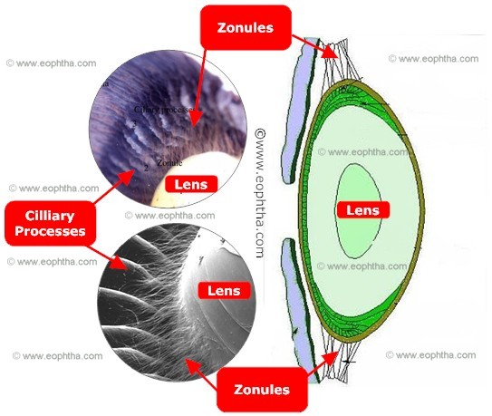
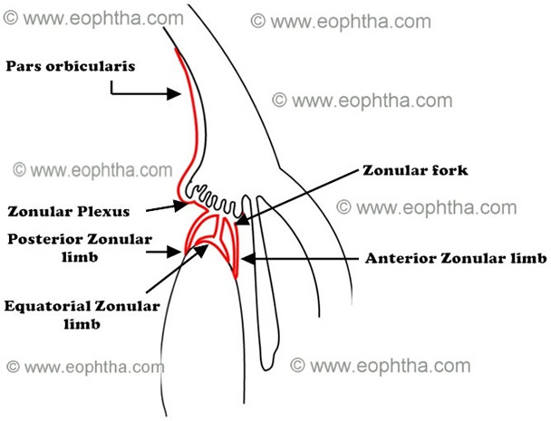
Most of the zonules arise from posterior part of parsplana, about 1.5 mm from ora serrate. Theses fibers run forward in pars plana blending with the basement membrane of the nonpigmented epithelium of pars plana. These fibers reach posterior margin of the pars plicata and turn into zonular plexuses. These zonular plexuses run through the valley of cilliary processes. Near anterior margin of the pars plicata, each zonular plexuses convert into zonular forks, which consist of three zonular fiber groups- anterior, posterior and equatorial fibers.
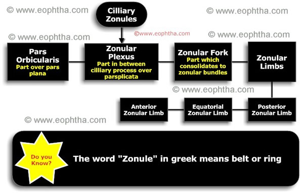
So, for the ease of description, the zonular apparatus of the eye can be divided into the following parts:
- Pars orbicularis: The part of the zonules which lie over pars plana.
- Zonular plexus: part of the zonules that lie between the cilliary processes.
- Zonular fork: the point of angulation of the zonule, which lies at the mid zone of cilliary valleys.
- Zonular limbs: consists of
- Anterior zonular limb:passes from pars plana to preequatorial part of the lens.
- Posterior zonular limb:passes from pars plicata to postequatorial part of the lens.
- Equatorial Zonular limb:passes from pars pliacata to lens equator.
Zonules are arranged in bundles which are composed of 2-5 fine fibers. Each zonular fiber is composed of multiple filaments of fibrillin, which are 8 to 12 nm in diameter.
Zonular fibers are rich in fibrillin, which maps to chromosome 15q.21.1.Mutations in fibrillin gene occur in Marfan’s syndrome, which causes weakening of zonules and subsequent subluxation of the lens.
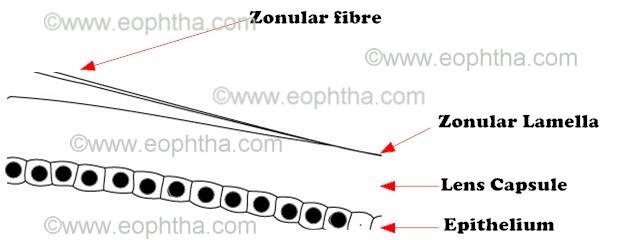
These zonules blend with the basement membrane of the lens capsule. This part of the lens capsule is known as zonular lamella and it is rich in glycosaminoglycans than rest of the capsule.
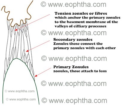
Most of the zonules attach to the preequatorial and postequatorial region of the lens capsule (approximately 1.5 mm from the equator) and few attach to the equator. The preequatorial zonules are relatively dense than the postequatorial zonules. Depending on their attachment, the zonules can be further divided into –
- Primary zonules:zonules, those attach to lens is called primary zonules.
- Secondary zonules:Zonules those connect the primary zonules with each other is called secondary zonules.
- Tension zonules or tension fibres:These are fibers which anchor the primary zonules to the basement membrane of the valleys of cilliary processes. They play an important role in accommodation.
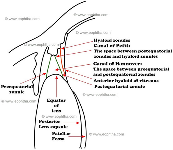
The space between preequatorial and postequatorial zonules is called Canal of Hannover. It is filled with the fine fibres of the equatorial zonules. The space between postequatorial zonules and hyaloid zonules are known as canal of Petit. Hyaloid zonules are the single layers of fibers which connect the anterior hyaloid of vitreous at the border of the patellar fossa to pars plana and pars plicata.