LASER is an acronym for “light amplification by stimulated emission of radiation”. Invented more than 50 years ago, today lasers are synonymous with precision and sophistication. The first medical application of lasers was the use in retinal photocoagulation and that role has been ever-evolving. Today its application is evident in all subspecialties of ophthalmology as a diagnostic and therapeutic tool.
Since medieval times, the damaging effects of strong light have been known to mankind. Socrates advocated avoiding direct viewing of the solar eclipse. The first documented use of light therapeutically was by Meyer- Schwickerath. He focused sunlight on the retinas of patients to treat melanomas. An undocumented early experiment with the aim of therapeutic photocoagulation was done by Moran- Salas in the 1940s. Meyer- Schwikerath highlighted that photic burns may be beneficial as a therapy. However, the major limitation of this was the requirement of enough sunlight to achieve the desired effect. This led to the evolution of non-solar sources of light like Carbon arcs, Xenon arc photocoagulators by Zeiss, etc. The first functioning LASER was produced by Mainman and colleagues.
What makes Laser radiation different from its counterparts like thermal radiation and other light radiation sources is the fact that the photons in a laser are coherent (same phase), monochromatic (narrow range of wavelength), and that it is a collimated beam (directional). The coherence of the laser beam is an important factor. The spatial coherence allows for small burns to the target tissue with minimum collateral damage, while the temporal coherence allows specific tissue targeting by selecting wavelengths that are absorbed only by the target tissues.
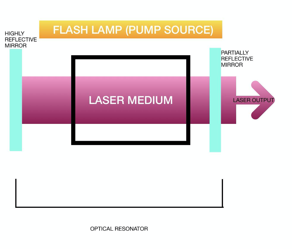
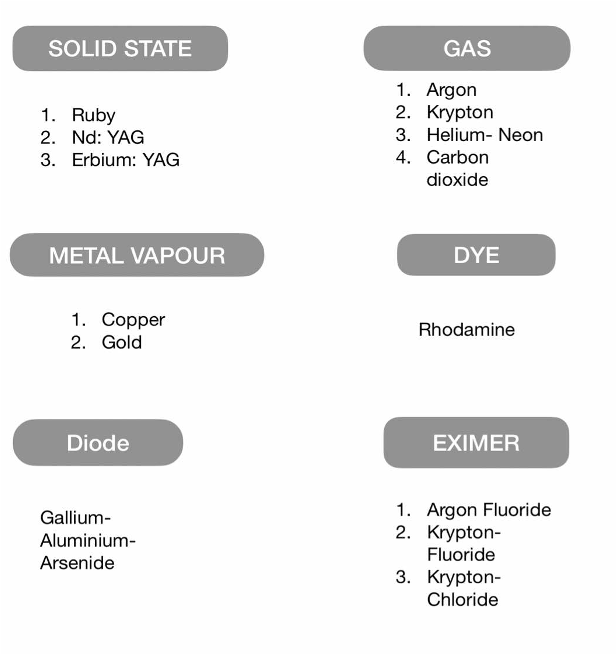
A typical laser system involves a gain medium, such as a ruby crystal or Nd: YAG (gain medium is a material that has the properties to amplify laser beam by stimulated emission), a reflective optical cavity, and a mechanism that excites electrons to higher energy states.
LASER- TissueInteraction:
Following are the various laser-tissue interactions:
A. PHOTOTHERMAL: This includes:
- Photocoagulation: the laser beam is absorbed by a target tissue, this results in the rise of temperature that causes denaturation of proteins. Eg: Argon, Krypton, Diode (810), and Frequency-doubled ND: YAG laser.
- Photovapourisation: thelaser beam is absorbed by the target tissue leading to the vaporization of the intracellular and extracellular water. This has specific uses in surgery as the adjacent blood vessels are also treated, thus avoiding spillage of blood. Eg: carbon dioxide laser with far-infrared wavelengths.
B. PHOTOCHEMICAL:This includes:
- Photoradiation: here a photosensitizing agent is injected intravenously to be taken up by target tissue. Thereafter the target tissue is exposed to red laser light. This generates cytotoxic free radicals and thus the desired laser effects. Eg: Photodynamic therapy.
- Photoablation: here laser in the far ultraviolet wavelength (<350 nm) is used. This results in the breakage of long-chain tissue polymers into smaller volatile fragments. Eg: Excimer laser
C. PHOTODISRUPTION (PHOTOIONIZING):Here laser is applied to the target tissue. The laser strips electrons from the target tissue. This creates a shock wave that disrupts the target tissue. Eg: ND: YAG laser.
|
LASER interaction |
Mechanism of Action |
Clinical Effect |
Example |
|
PHOTOTHERMAL (Photocoagulation and Photovapourisation) |
Coagulation of proteins |
Tissue burns |
Argon, Nd: YAG laser |
|
PHOTOABLATION |
Breakage of interatomic bonds |
Tissue etching |
Excimer |
|
PHOTORADIATION |
Generation of cytotoxic free radicals |
Oxidative tissue damage |
Photodynamic therapy |
|
PHOTODISRUPTION |
Stripping of electrons from atoms, the formation of plasma/ shock wave |
Cuts tissues |
Nd:YAG laser (Q switched) |
LASER DELIVERY SYSTEMS:
Following are the techniques for delivering laser to the target tissues in ophthalmology:
- Slit-lamp: This is a common mode of delivery of laser to the target tissue where the power, spot size, etc can be easily modulated.
- Indirect ophthalmoscope: a fiber optic cable delivers diode or argon laser to the retina. Spot size depends on the diopteric power of the condensing lens used. Eg: treatment of peripheral retinal breaks.
- Endophotocoagulation: this system is used at the time of surgery via pars plana route as in vitrectomies or treatment of proliferative diabetic retinopathy. A fiber optical cable delivers argon green or diode laser.
Uses of LASER in Ophthalmology:
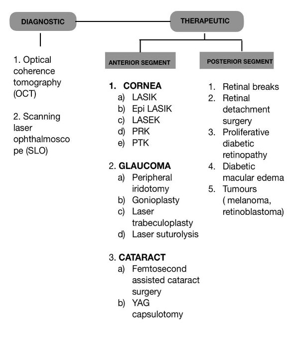
Lasers in ophthalmology have a diagnostic and therapeutic role. The flow chart below gives a brief summary of the uses of laser in ophthalmology.
A) DIAGNOSTIC USES: laser technology has been utilized in investigation techniques such as Confocal scanning laser ophthalmoscope (CSLO), which is used for optic nerve head evaluation, Optical coherence tomography (OCT), which is useful in the evaluation of retinal pathology, corneal pathologies, etc. Laser retinal Doppler flowmetry is useful in evaluating the optic nerve head perfusion.
|
ANATOMICAL SITE |
LASER PROCEDURE |
INDICATION |
TYPE OF LASER |
|
IRIS |
Peripheral iridotomy (PI) |
Angle closure glaucoma Occludable angles |
Nd:YAG or Argon- YAG |
|
Laser iridoplasty |
Plateau iris Unresponsive angle closure glaucoma Prior to ALT |
Argon |
|
|
Laser pupilloplasty |
Breaking peripheral anterior synaechiae (PAS) Relieve appositional angle closure Adjust the position of a decentred pupil |
Argon |
|
|
SCLERA |
Laser scleroplasty |
Primary open angle glaucoma (POAG) |
Holmium YAG |
|
Laser suture lysis |
After trabeculectomy to increase filtration |
||
|
ANGLE |
Laser trabeculoplasty (ALT) |
Refractory POAG Pigment dispersion Pseudoexfoliation syndrome |
Argon |
|
Selective laser trabeculoplasty (SLT) |
Argon |
||
|
CILIARY BODY |
Cyclophotocoagulation (CPC : transscleral/ trans pupillary/ endoscopic) |
Refractory glaucoma like: Neovascular glaucoma (NVG) Uveitis glaucoma Traumatic glaucoma Congenital glaucoma |
Diode Nd: YAG |
|
ANTERIOR VITREOUS FACE |
Laser vitreolysis |
Malignant glaucoma |
Nd: YAG |
B) THERAPEUTIC USES:
ANTERIOR SEGMENT:
1. CORNEA: Lasers have been utilized in procedures for refractive error correction like LASIK, epi-LASIK, LASEK, PRK, and therapeutic procedures such as phototherapeutic keratectomy (PTK) for the removal of surface irregularities, small corneal opacities.
2. GLAUCOMA: Thefollowing are the therapeutic applications of laser in glaucoma depending on the anatomical site
3. CATARACT: Thelaser is utilized to assist cataract surgeries like Femto laser-assisted cataract surgeries (FLACS) and post-cataract surgery for removal of capsular opacification as in Nd: YAG posterior capsulotomy.
POSTERIOR SEGMENT:
RETINAL NEOVASCULAR DISEASES:
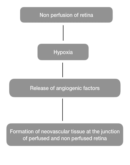
These include Diabetic retinopathy, Central and Branch vein occlusion, Sickle cell retinopathy, etc. PATHOGENESIS: The aim of laser treatment in these cases is to eliminate hypoxia retina so as to eliminate the stimulus for neovascularization. This is called Pan retinal photocoagulation. Argon or Krypton laser may be used for the same
2. RETINAL TEARS AND RETINAL DETACHMENT:
Retinal tears and small retinal detachments may be treated with Focal laser photocoagulation. Here many small burns are applied to the attached retina around the tear or localized detachment in an attempt to “weld” the retina.
3. CHOROIDAL NEOVASCULAR DISEASES:
Conditions such as age-related macular degeneration, choroidal neovascularization secondary to trauma, angioid streaks, etc can be treated with Argon or krypton laser photocoagulation
4. INTRAOCULAR TUMORS:
Tumors like Retinoblastomas, melanomas, and retinal angiomas can be treated with focal Argon or Krypton lasers.
5. ANOMALOUS BLOOD VESSELS AND MACULAR EDEMA:
In conditions like Retinal microaneurysms and Coats’ disease, direct focal treatment of the vessels may lead to resolution of the condition. Leaking vessels may lead to macular edema. The treatment of such vessels with a focal laser may help manage the associated macular edema.