
Introduction
The eyeball development begins early in the 4th week of intrauterine life with formation of optic vesicle (a diverticulum) from diencephalon. The structure of eyeball is shown in Figure 1. Various components of eyeball are derived from the following sources:
- Retina, iris and optic nerves are derived from optic vesicle that arises from neuroectoderm of diencephalon.
- Lens and corneal epithelium are derived from lens placode that arises from surface ectoderm.
- Fibrous and vascular coats of eyeball are derived from mesodermal condensation surrounding the optic vesicle.
- Choroid and sclera are derived from migrating neural crest cells.
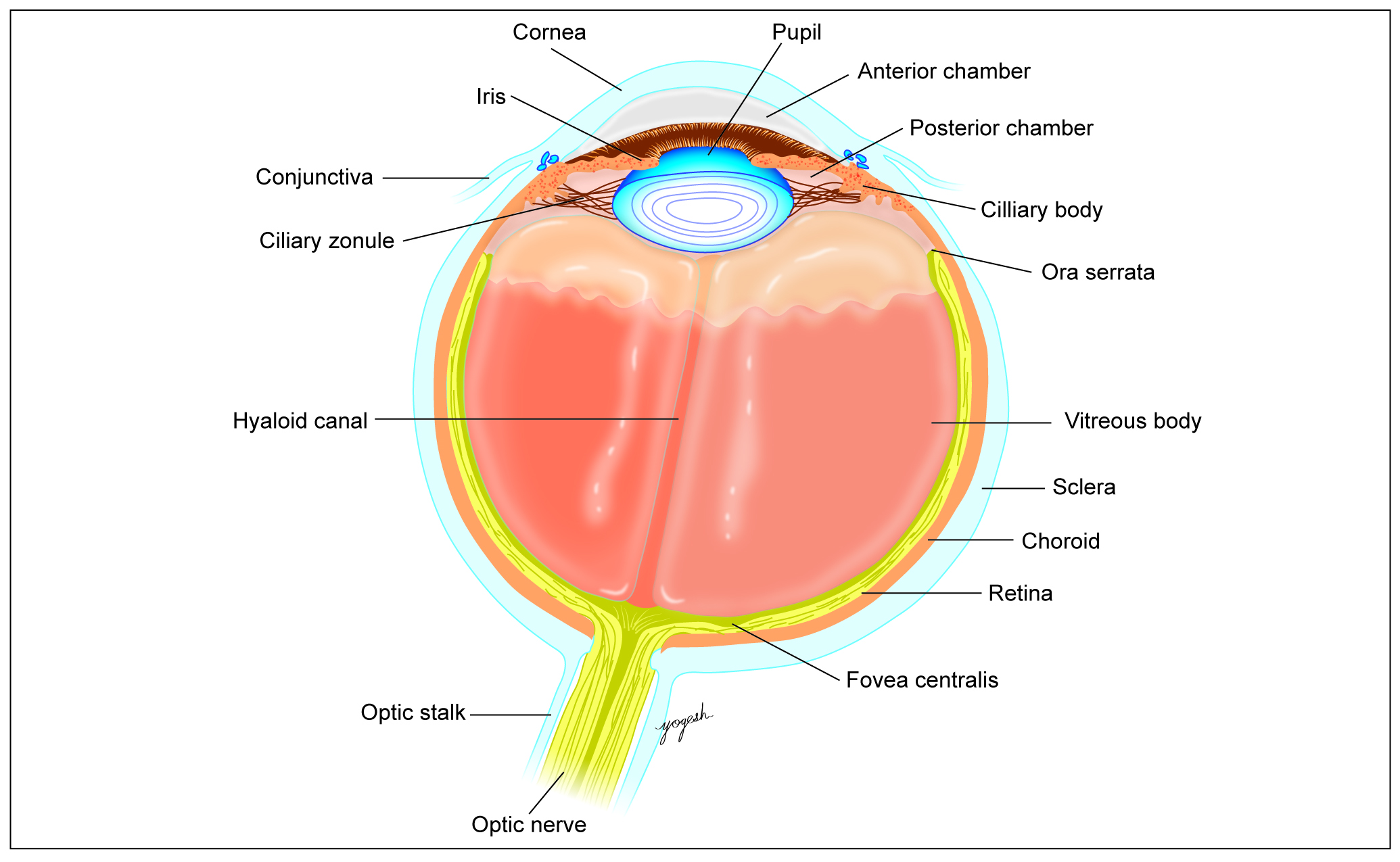
Figure 1: Fully developed eyeball
Optic Vesicle and Lens Vesicle
Formation of optic sulcus: On 22nd day, wall of the diencephalon shows thickening and depression to form optic sulcus. Formation of the optic sulcus or groove is the first indication for development of the eye (Figure 2A). Formation of optic vesicle: Optic sulcus further invaginates laterally in the surrounding mesoderm and form a bulging called optic vesicle (Figure 2B). Formation of optic stalk: Optic vesicles grow laterally but remain connected to forebrain by a stalk-like structure called optic stalk (Figure 2B). Formation of lens placode: Optic vesicle meets surface ectoderm. In region of contact, the surface ectoderm forms localized thickening called lens placode (Figure 2B). Formation of lens vesicle: Lens placode depresses to form lens pit and later, lens vesicle (Figure 3A). Lens vesicle loses contact with surface ectoderm by 33rd day. Formation of optic cup: Lens vesicle invaginates the optic vesicle and converts it to a double layered optic cup (Figure 3B). By 6th week, margins of the optic cup overgrow and cover lens vesicle except on caudal side of the lens and partly on caudal surface of optic stalk. This gap is called choroidal fissure or foetal fissure (Figure 3B). In a mesodermal encroachment along the choroidal fissure, hyaloid vessels develop that supply optic and lens vesicles. The distal part of hyaloid vesicles degenerates, whereas the proximal parts from central artery of retina and central vein of retina (Figure 4).
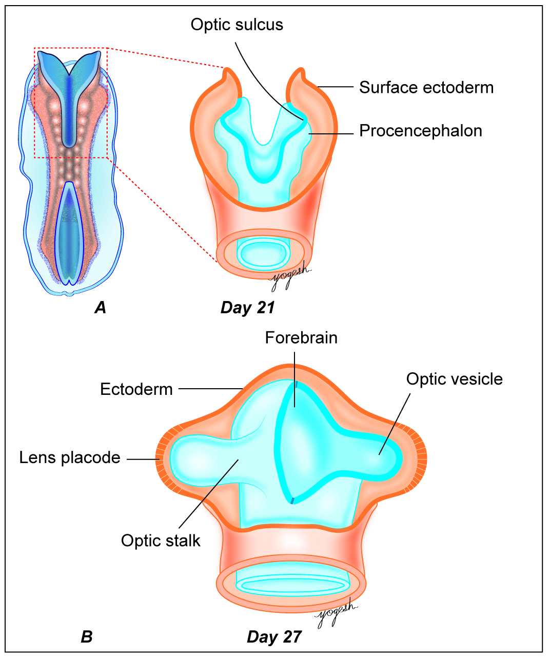
Figure 2: Development of eyeball on day 21 and 27
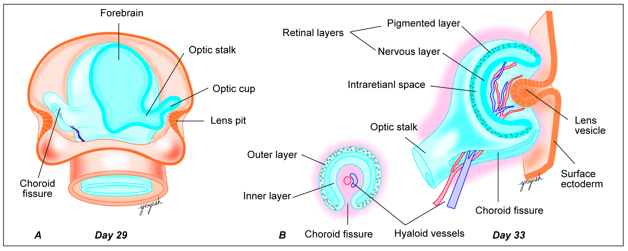
Figure 3: Further development of eyeball (day 29 and day 33)
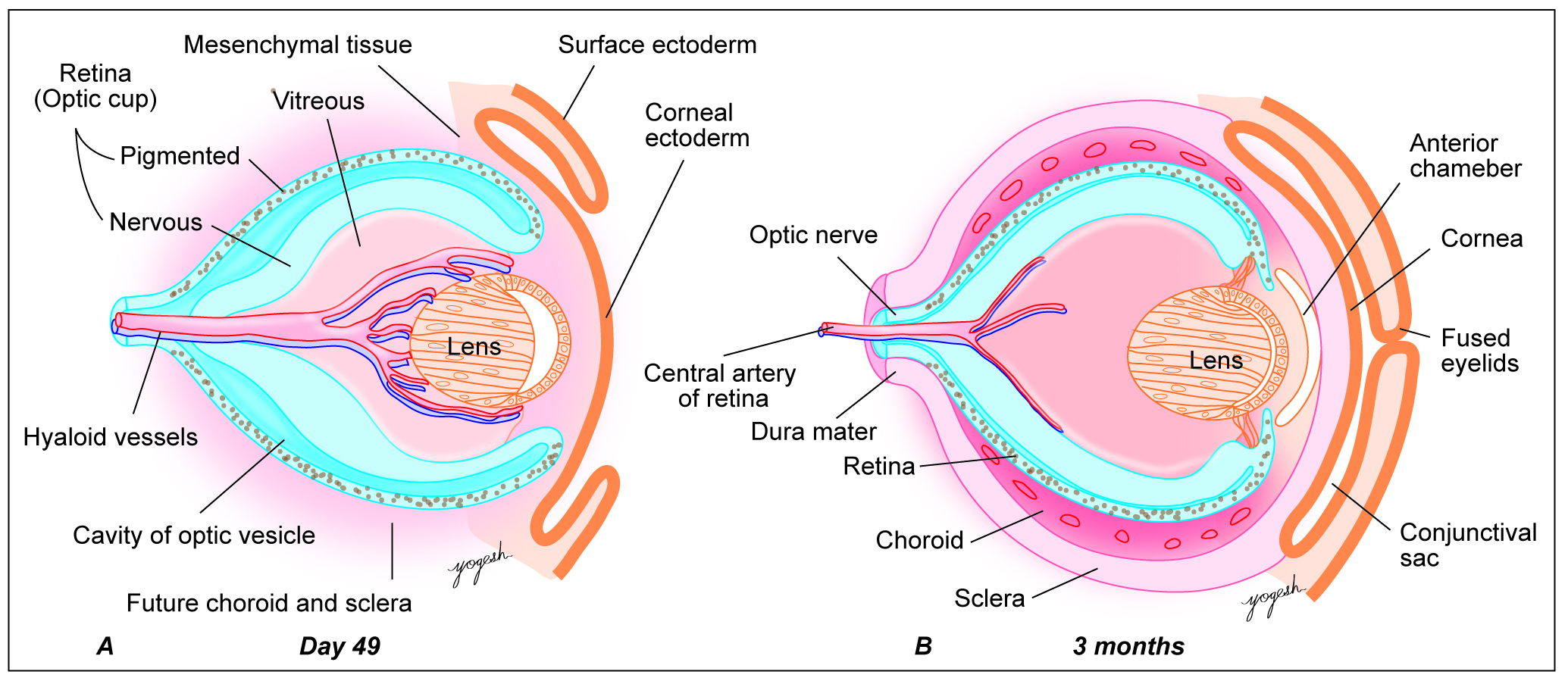
Figure 4: Further development of eyeball (day 49 and 3 months)
DEVELOPMENT OF VARIOUS PARTS OF EYEBALL
Cornea
Cornea consists of outer stratified squamous epithelium resting on basement membrane, lamina propria, Descemet’s membrane and inner corneal epithelium. All these layers are derived from the following sources:
- Surface ectoderm forms outer stratified squamous epithelium and its basement membrane (Bowman’s layer).
- Lamina propria or stroma is derived from mesoderm.
- Neural crest cells form Descemet’s membrane and inner corneal epithelium.
Lens
Lens is a transparent, biconvex structure that lies between aqueous and vitreous of the eyeball. Lens has three main parts:
- Lens capsule
- Lens epithelium (simple cuboidal epithelium)
- Lens fibres
Stages of Development
By 33rd day of IUL, lens vesicle gets separated from surface ectoderm. Initially lens vesicle is lined by a single layer of cuboidal epithelium (Figure 5D). Cells of ventral wall of lens vesicle remain cuboidal. Cells of deep wall of lens vesicle elongate, become columnar and occupy cavity of the lens vesicle (Figure 5E). These elongated cells of posterior wall lose their nuclei and form transparent primary lens fibres (Figure 5F). The equatorial cells continue to contribute new lens fibres and lens grow. These fibres later become hard and form secondary lens fibres. Cells of anterior wall persist and form epithelium of lens. On degeneration of hyaloid artery (that supplies lens), lens becomes avascular structure. In 7th week, solid lens is formed.
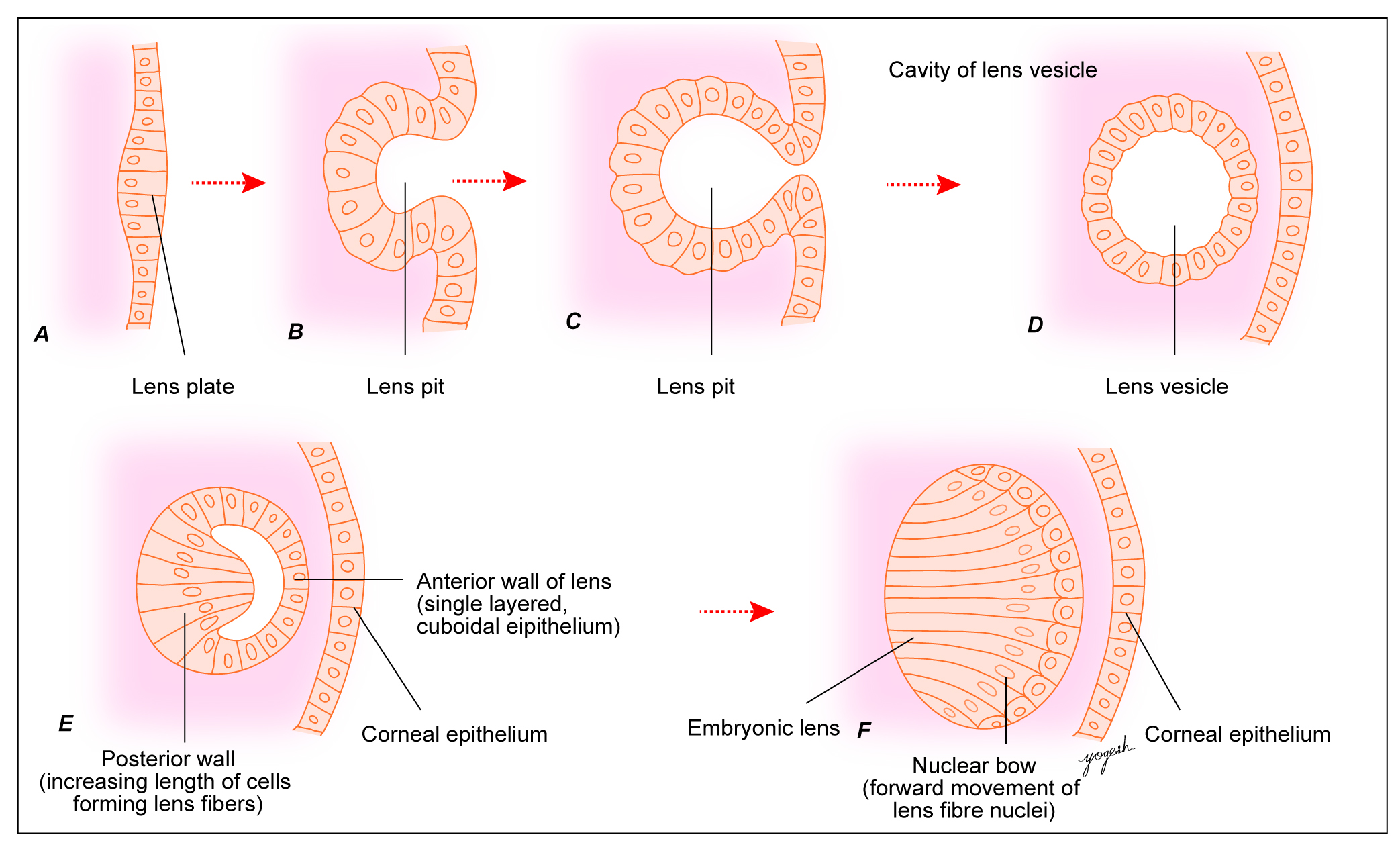
Figure 5: Development of the embryonic lens
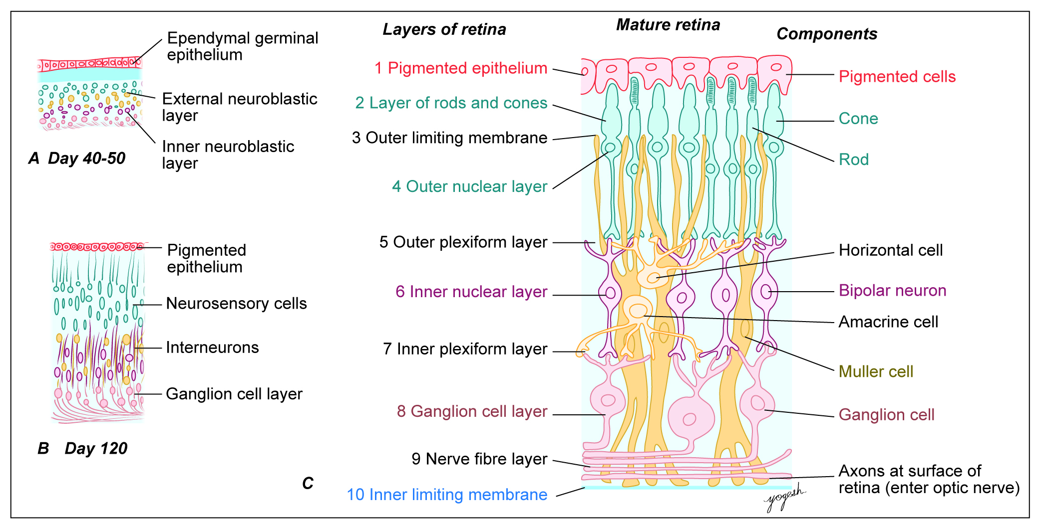
Figure 6: Histogenesis of retina
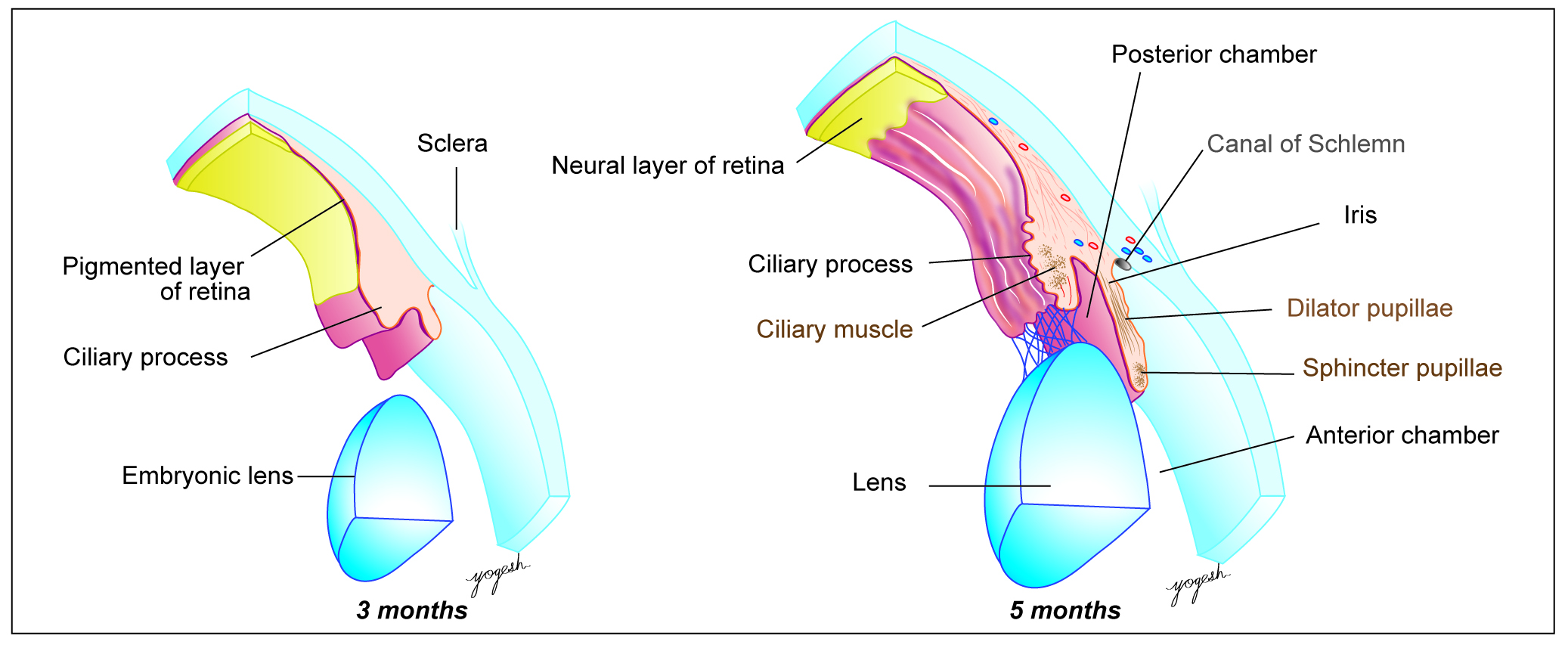
Figure 7: Development of the iris and ciliary body
Retina
Cellular layers of retina from outside inwards include:
- Pigmented cell layer
- Layer of rods and cones
- Bipolar neuron layer
- Ganglionic cell layer
All these layers are derived from the optic cup.
Stages of Development
From wall of diencephalon, optic vesicle arises that grows laterally but remains connected by optic stalk with the diencephalon (Figure 2B). Optic vesicle comes in contact with lens vesicle and gets converted to optic cup (Figure 3). Optic cup has two parts, anterior and posterior. Anterior part of optic cup forms an epithelial covering of ciliary body and iris. Posterior part of optic cup forms various layers of retina as follows:
– Outer wall: Forms pigmented layer of retina.
– Inner wall: Gets differentiated into three layers as
- Matrix layer – forms rods and cones.
- Mantle layer – forms bipolar cells and ganglionic cells as well as other neurons of retina.
- Marginal layer – forms optic nerve. Axons of ganglion cells form marginal layer that converges towards optic stalk and finally, forms optic nerve.
Space between outer and inner wall of the optic cup is called intraretinal space. This space gets obliterated by increasing number of retinal neurons.
Optic Nerve
Optic stalk contains hyaloid artery and vein, axons of ganglionic cell layer. On inferior (caudal) surface of optic stalk, choroid fissure is present, that closes by 7th week. Later, optic stalk forms optic nerve, whereas hyaloid vessels form central artery and vein of retina.
Congenital Retinal Detachment
It involves separation of pigmented epithelium from neuronal layers of the retina. Congenital retinal detachment mostly occurs as an autosomal recessive disorder. It results in permanent blindness from birth.
Sclera and Choroid
Sclera (white of eye) is opaque, fibrous protective outermost layer of eyeball. Choroid is a vascular coat of eyeball that lies between outer sclera and inner retina. During the 6th and 7th weeks, mesenchyme that surrounds external surface of optic cup condenses into two layers: Outer fibrous layer that forms sclera, Inner vascular layer that forms choroid. Choroid is anteriorly continuous with ciliary body and posteriorly with pia and arachnoid mater. Sclera is anteriorly continuous with substantia propria or stroma of the cornea and posteriorly with dura mater covering optic nerve.
Ciliary Body
Ciliary body is a ring-shaped mass that divides eyeball separates aqueous humour from vitreous. It consists of ciliary muscle that controls shape of the lens. Ciliary body shows folding of epithelium called ciliary processes that secret the aqueous humour. Mesoderm surrounding the anterior part of optic cup forms ciliary muscle and connective tissues of ciliary body. Pigmented epithelial lining of inner aspect of ciliary body and ciliary processes is derived from outer layer of the optic cup; hence, continuous with pigmented layer of retina. The nonpigmented layer of epithelium covering deeper side of ciliary body is derived from inner layer of optic cup and continuous with neuronal layers of retina.
Iris
Iris is a thin, circular structure in eyeball that controls size of the pupil. Iris shows anterior limiting layer, stroma, sphincter and dilator papillae muscles and posterior pigmented epithelium. Iris develops from anterior extension of optic cup. Epithelium (anterior and posterior) of iris is derived from double layered optic cup. Neuroectodermal cells of optic cup also give rise to muscles of the iris. Stoma of the iris is derived from neural crest cells. Colour of eye/pigmented layer of cornea depends on genetic constitution of an individual.
Anterior and Posterior Chambers of Eye
Anterior and posterior chamber is similar to subarachnoid space of brain. Mesoderm located between lens and cornea split to form spaces. These spaces split into anterior and posterior chambers by developing iris and pupillary membrane. As pupillary membrane disappears, anterior chamber communicates with posterior chamber through pupil. Epithelium of ciliary process starts secreting aqueous humour that fills anterior and posterior chambers of eyeball. Sclera of sinus venosus forms a network of trabecular meshwork at sclerocorneal junction along the circumference of anterior chamber. These later form canal of Schlemm and drains aqueous humour. [Canal is named after Friedrich Schlemm (1795–1858), a German anatomist.]
Vitreous
Vitreous occupies chamber of eyeball called vitreous chamber. Vitreous is a transparent, gelatinous mass that forms about 45th volume of the eyeball. Vitreous develops as follows: Primary vitreous humour develops from neural crest cells of optic cup. Secondary vitreous humour replaces the primary vitreous. Secondary vitreous humour is derived from inner layer of optic cup and lens vesicle. Vitreous humour is supplied by hyaloid artery that later gets obliterated.
Accessory Structures of Eyeball
Extraocular Muscles
Each eyeball has 6 extraocular muscles, namely superior rectus, inferior rectus, medial rectus, lateral rectus, superior oblique and inferior oblique muscles (levator palpebrae superioris muscle for eyelid). All extraocular muscles are derived from preoccipital myotomes and hence, supplied by 3rd, 4th and 6th cranial nerves.
Eyelids and conjunctival sac
Eyelids are developed by folds of surface ectoderm that raise above and below the cornea. These ectodermal folds contain some mesoderm that gives raise to tarsal plate and other connective tissue of eyelids. Eyelashes and glands of eyelids develop from surface ectoderm. Surface ectoderm that lines inner surface of eyelids and covers eyeball form conjunctival sac. Eyelids remain fused till 28th week of IUL and later get separated.
Lacrimal apparatus
It consists of lacrimal gland, lacrimal sac and nasolacrimal ducts. Lacrimal gland is derived from 15 to 20 buds that grow at superolateral angle of conjunctival sac. Nasolacrimal duct and lacrimal sac develop from naso-optic furrow (nasolacrimal furrow). Lacrimal canaliculi develop from canalization of ectodermal buds that raise from medial margins of eyelids. Later these canaliculi develop communication with lacrimal sac.
Congenital Anomalies of Eye
- Anophthalmia is an absence of eyeball due to failure of optic vesicle formation.
- Microphthalmia is small eye due to underdeveloped optic vesicle. It may be due to rubella virus, cytomegalovirus, toxoplasma or other intrauterine infections. PAX6 gene performs key-regulatory role in the development of eye. PAX6 gene mutation results in aniridia or microphthalmia.
- Cyclopia is a presence of single median eye. It occurs due to fusion of two optic vesicles (synophthalmia). Single eye may be connected with face by a tubular stalk called proboscis. Inhibition of sonic hedgehog (SHH) gene may result in Cyclops.
- Coloboma of iris: Due to failure of fusion of choroid fissure, iris shows a cleft on inferior side (keyhole appearance). This condition is called coloboma of iris (coloboma = missing part, Greek).
- Persistent papillary membrane: Pupil may be covered partially or completely by persistent papillary membrane.
- Congenital aniridia is a complete absence of iris due to an arrest of development of optic cup.
- Congenital aphakia is an absence of the lens of eyeball due to failure of formation of lens vesicle.
- Coloboma of eyelid is an absence of part of the eyelid. Mutation of PAX2 gene results in optic nerve colobomas and renal aplasia.
- Entropion is inward-turned eyelids, whereas ectropion is outward turned eyelids.
- Epicanthus is a crescentic fold of skin that extends from upper eyelid to canthus. It is a feature of Mongolian races and Down syndrome (trisomy 21).
- Cryptophthalmos is an absence of eyelids and palpebral fissures. The eyeball is covered by skin.
- Congenital ptosis (drooping of the eyelid) occurs due to the failure of the development of levator palpebrae superioris muscle.
 Source: Textbook of Human Embryology, Yogesh Sontakke, CBSPD, Delhi, India.
Acknowledgment: CBS Publishers and Distributors Pvt Ltd, Delhi, India.
Copyright: Dr. Yogesh Sontakke and CBS Publishers and Distributors Pvt Ltd, Delhi, India.
Source: Textbook of Human Embryology, Yogesh Sontakke, CBSPD, Delhi, India.
Acknowledgment: CBS Publishers and Distributors Pvt Ltd, Delhi, India.
Copyright: Dr. Yogesh Sontakke and CBS Publishers and Distributors Pvt Ltd, Delhi, India.