

An operating surgical microscope is the most important optical instrument in the modern era of ophthalmic surgeries. It provides the surgeon with a magnified and illuminated high-quality image of the small ophthalmic structures. Being binocular the surgical microscopes gives the additional benefit of high-quality stereoscopy.
History
The first German doctor to use a stereomicroscope was Dr. Hans Hinselmann (1884-1959), Prof. of Gynaecology under Dr. Otto von Franque at the University Clinic, Bonn, who adapted in 1925 a Leitz stereomicroscope with coaxial illumination to a movable floor stand. He named IT “colposcope” and used it to visualize the female genital organs particularly for cancer of the cervix. Dr. Hans Littmann (1908 – 1991), head of Med-Lab at Zeiss OptonOberkochen, West Germany (now Carl Zeiss A.G.) and his team of technicians designed in 1953 their first ophthalmic operating microscope
Prerequisites for an ideal operating microscope
- Must have stereoscopic visualization.
- Must have a comparatively long working distance
- The illuminating system must evenly illuminate all the parts of the surgical field
- Stable with articulated balance.
- Possibility to adapt to a wide range of procedures.
Principle
The basic principle of the microscope is two convex lenses ( objective lens and eyepiece lens) separated to a certain distance more than the focal length of each lens. The primary requisite for the good ophthalmic microscope is smooth magnification without any change in working distance. Working distance is the distance between the objective lens and the main focusing point. For the smooth transition of this magnification, the operating microscope is fitted with a magnification changer based on the principle of the Galilean telescope.
Magnification The magnification of an image is a relative value and has to do with the size of an image as projected on to the retina of the eye (or onto a piece of film in the case of a camera). With the use of the optical aid, and without changing the distance value, the size of the image of an object can be increased on the retina. The formula to find out original magnification of an operating microscope is
The focal length of tubes/ Focal length of objective lens X Eyepiece X Magnification value =Total magnification
The optical aid increases the image size on the retina without the need to reduce the eye-to-object distance to get the desired effect as shown in figure 1
Parts of an Operating Microscope
The ophthalmic surgical microscope has 3 primary parts-
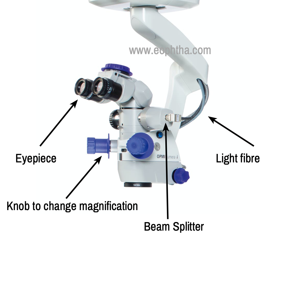
Optical system
The optical component consists of a basic stereo microscope with a binocular head, magnification changer, and an objective lens.The binocular head consists of two telescopes with the adjustable eyepiece. The telescopes can be adjusted for surgeons with refractive error. In the newer variants of microscopes, the telescope has 125mm of focal length. The machine has a knob in the microscope to change the magnification stepwise. Different desired magnification factors are engraved along the knob. Few higher-end machines have a continuous zooming system available in the foot paddle. Most of the microscope of the modern era comes with side-viewing scope for the assistant. A beam splitter is used for proper functionality and simultaneous viewing from the side scope.
Illumination system
In an ophthalmological surgical operating microscope the optical system generally vertically aligned but the illuminating system is obliquely placed. Arc shaped halogen light is mounted in the stand and the light is delivered to the optical system with a fiber optic wire to reduce the heat generation by the illumination system. The microscope illuminator transmits light to illuminate the surgical area. This light can be varied, just change the voltage to the light bulb. Most microscope floor stand power supplies have a provision to vary the light intensity by this method. Under the microscope, a specific amount of light will be projected and any change made in microscope magnification will have no effect on the amount of light being projected from the microscope.
The light in ophthalmic microscopes is usually co-axial, which means the light follows back the exact same path as the images in order to remove the shadowing. Coaxial illumination microscope illumination can be of two varieties.
Oblique illumination: Originally, microscopes had only externally mounted independent illuminators transmitting light but creating some shadows and unable to get down deep into cavities. The need to get the light into the neuro-surgical cavities, through ear specula and down into throats caused the development of the built-in microscope coaxial illuminator. 1
Coaxial type illuminator: the light from the illuminator bulb is re-routed to a point very near the viewing axis of the microscope and is projected down through the same objective lens used for viewing. In recent years, coaxial illumination has become standard on all ophthalmic microscopes.
Working Distance: The working distance is the distance from the microscope objective lens to the point of focus of the optical system. This value is fixed and is dependent on the chosen focal length of the objective lens. The choice of working distance depends on the type of surgery. For modern ophthalmic surgery that involves delicate work in the posterior chamber, objective focal lengths of 150 mm, 175 mm and 200 mm are commonly used.
Par-FocalityThis term refers to the state of optical configuration that enables magnification changes to take place without affecting the point of microscope focus.
Depth-of-Field / Depth-of-Focus These terms relate to the area in front of, and behind, the point of perfect optical focus, where sharp focus is maintained. Higher the magnification, the more shallow the marginal area of sharp focus and vice-versa.
Resolution This optical term, simply stated, is the quality of a lens (prism or mirror) which enables it to deliver a perfectly crisp image where intended. Resolution and depth-of-field are reciprocals of one another in that more resolution implies less depth-of-field in any one optical design
Magnification Changer It is a method for varying magnification of a basic optical system. It may consist of one or several supplemental lens elements that are moved in and out of the viewing axis, or it may be a system of shifting lens elements which change their positions relative to one another such as in a zoom magnification system. (Figure 2)
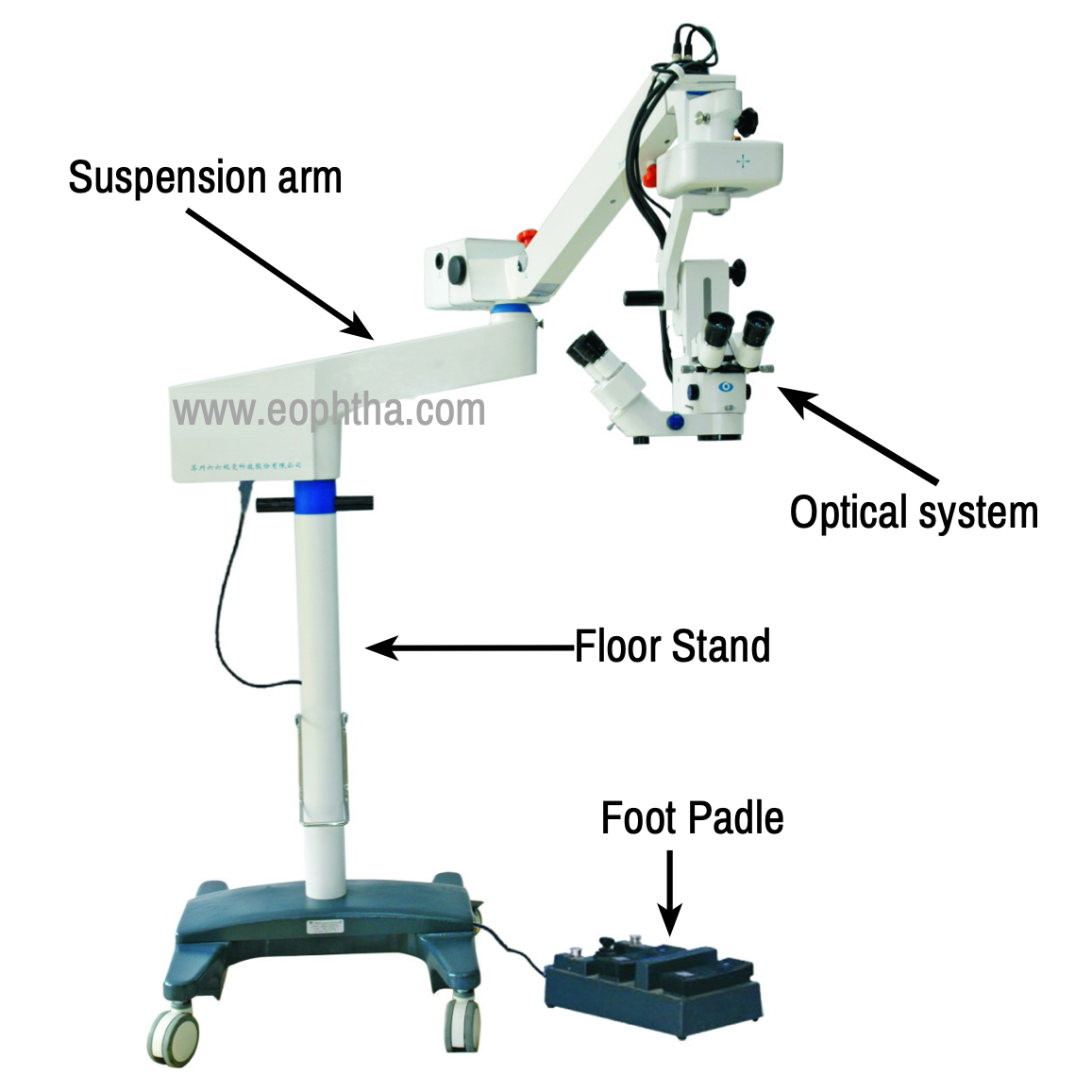
Stand / Suspension Arm
The whole optical system stands stable from the floor through the stand and suspension arm. The stand has wheels and can be moved around and can be fixed with brakes. The suspension arm makes it possible to focus it to focus at the exact point of interest with comfort to the surgeon. The microscope optical system is suspended through the gimbal technique which makes the system “weightless” leading to no additional momentum to any directions when moved in any directions (FIG:3). The stands are often connected to foot paddle for controlling the focus and zoom. The control joystick attached in the foot paddle and translate the optical system in the horizontal axis also (X,Y). Ophthalmic microscopes are the most important tool in the current era where we are perusing all the surgeries for better visual acuity and lesser postoperative astigmatism requiring more finer visualization of the microscopic structures.
It is very important to learn how to operate the microscope before your first day in the OPERATING THEATRE
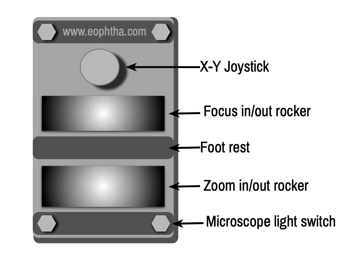
Basics of the Microscope pedal. While there are some subtle variations among models and manufacturers the basics of the microscope foot pedal and operation of the microscope are similar. The microscope has a starting XY position which is centeredat the start of the case and then small variations in this initial position are made using the foot pedal which makes small XY adjustments of the microscope.Often the intensity of the light can also be controlled with the foot pedal.
Allof your extremities will be busy:one foot for the microscope pedal, one foot for the phaco foot pedal, one hand for the phaco-hand piece, and the other hand for the chopper. Most surgeons use the non-dominant foot to control themicroscope pedal.Most people take off their shoes so that they can feel the microscope pedal better.
Foot pedal switches The typical positions of the microscopes footswitch controls are shown below. The foot pedal is designed so that the foot can sit on a raised footrest. A rocker switch in front of the footrest is most important and moves the scope up and down to make small changes in focus. a rocker switch behind the footrest controls the zoom or magnification. The magnification is typically low duringwound construction and is increased during steps such as capsulorhexis which require more magnification.Several inches in front of the footrest is thejoystick which controls the XY position of the scope. Both the XY position and the focus should be centered prior to the case (usually a switch on the scope) and manually put into the optimal initial position to allow the maximal excursion of these functions during the case.
What’s New
Alcon:Alcon’s new addition, the LuxorLX3 with Q-VUE Ophthalmic Microscope, offers an expanded visual field, a large and stable red reflex, and excellent visual detail.The red reflex capabilities sound promising, featuring a technology called ILLUMIN-i to provide a large, stable red reflex zone “regardless of pupil size, centration, eye tilt or patient movement.”
TrueVision enables the surgeon to operate in a heads-up manner by 3D viewing a large screen instead of traditional microscope oculars
The Luxor is part of Alcon’s Cataract Refractive Suite, an interconnected group of devices that also includes the Verion Image Guided System, Centurion phaco machine, LenSx femtosecond cataract laser, and ORA VerifEye+ aberrometer, as well as an integrated HD video system.
OPMI Lumera 700 Ophthalmic surgical microscope from Zeiss features Stereo Coaxial Illumination (SCI) for a highly stable and high contrast red reflex. Increase workflow efficiency with markerless toric IOL alignment Cockpit integrates data from all connected devices, including VISALIS 500 from ZEISS.
Optimize light intensity, magnification and centration of the live image with the guided image quality check for retinal surgeons, the OPMI Lumera 700 and Resight 700 fundus viewing system allow recognition of every detail of the retina. It can be equipped with the Rescan 700, a fully integrated intraoperative OCT, which projects real-time, high-definition OCT images into the eyepiece for both anterior and posterior segment procedure
Haag-Streit Surgical The surgical division of Haag-Streit offers the Hi-R NEO 900 for anterior and posterior surgery. The microscope has HD video capabilities for both 2D and 3D, integrated OCT (iOCT), and the integrated ophthalmoscope EIBOS 2 system for retina surgery. The Hi-R NEO 900 can incorporate the Sony 3D viewing system or the TrueVision system.