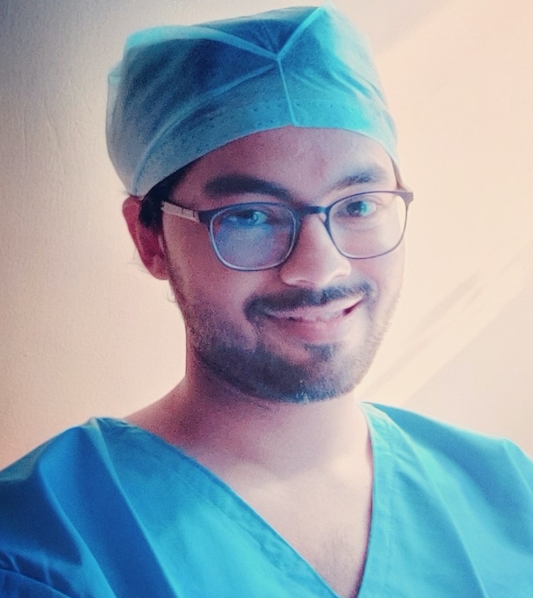
 Retinal imaging has come a long way today. From times of appropriately concentrating the fluorescein dye from scratch in the lab itself to times where we have a dye-free angiography system, technology has covered vast boundaries. There is a platter of different modalities in the market today, each having their own ebb and flow. And what the future beholds for us is certainly inexplicable. To throw a light on these modalities, we have a special interview with someone who continues to extensively work on these technologies and modalities day in and out,Dr. Manish Nagpal
Dr. Nagpalis practicing asa vitreoretinal consultant at Retina Foundation, Ahmedabad, since 1999. He completed his Ophthalmology training at M & J Institute of Ophthalmology, Ahmedabad in 1996 and further went on to accomplish the FRCS (Ed) at the UK in 1997. He is a recipient of the prestigious Distinguished Service Award of APAO in 2007, the Achievement award of APAO in 2012, the American Academy’s Achievement award 2009 as well as AAO ‘sInternational Ophthalmologist Education Award in 2011, and the AAO Senior Achievement Award in 2018He has a keen interest in making surgical videos and has received the prestigious Rhett Buckler award for best video at the annual meeting of ASRS (5 years in a row)2009New York,2010Vancouver,2011 Boston followed by Las Vegas, 2012 and Toronto, 2013.He received the Best Video award at the film festival held at the 25thAPAO meet held at Beijing, China, in Sept 2010 and the Best of Show at the annual AAO meeting held at New Orleans in Oct 2013. At the annual All India congress ( AIOS) he has won the best paper in Retina four times including the S Natarajan award in 2008 and the C S Reshmi award for the best video presentation in 2012. He is also the recipient of the IIRSI special Gold medal for 2012 as well as the Young achievers' award from BOA in 2012. He is also the recipient of the Honor award @ 2013 and the Senior Honor award in 2014 for outstanding contribution to the American Society of Retinal Surgeons (ASRS).He is also the recipient of the (Indian journal of Ophthalmology) IJO Gold award for 2014. He has numerous peer-reviewed articles to his credit along with numerous chapters in various international books. He is a regular reviewer with ophthalmology journals such as RETINA, EYE, European Journal of Ophthalmology, and IJO. He is part of the select Program Committee for The Prestigious Vail Vitrectomy meeting held in 2016 and 2019. He has been part of the Florentina program committee in 2015 and was invited to perform live surgery from Athens to be transmitted to Florence for the Florentina, 2019.He ison the international editorial boardofRETINA TODAYand Ocular surgery news (OSN). He has been invited by ORBIS for a Voluntary surgical program on the Flying Eye Hospital (FEH) at Chittagong, Bangladesh in 2017 and works closely with ORBIS for their online forum Cybersight for surgical videos and web-based presentations. He has conducted various Vitreoretinal surgical workshops doing live surgery and laser procedures at various meetings in India and abroad. He has beena Principle investigator for manyglobal multicentric trials including (Macugen 1013, VIEW 2, DEGAS, and MONET).
eOphtha: There is wide discussion on OCT Angiography (OCT-A) replacing FFA in the field of medical retina in the near future. What is your take on that? How do you compare the clinical output of OCT+FFA vs OCT-A, if we place the non-invasiveness and repeatability perks of the latter aside?
Dr. Manish Nagpal: OCT Angiography is certainly a revolutionary imaging technology. It is still in its native phase and needs to stand test of time since OCT and FA have long been sub-serving the purpose. A common vocabulary needs to be developed among the interpreters worldwide for its acceptability. Undoubtedly, it scores a lot in terms of non-invasive nature and ability to provide accurate information on repeated examinations. Some of the data in terms of interpretation of morphology of pathologies, exact location, in depth analysis, identification in terms of vasculature, blood flow measurements etc. that is provided by OCT angiography makes it more deserving. In current scenario due to variation in different OCT angiography machines and the fact that it needs to stand test of time for reliable results and for a common interpretation guideline along with high cost does not make it a tool that will replace OCT and FFA in near future. Moreover, peripheral examinations on OCTA are still limited and has a long way to go
eOphtha: How do we differentiate a Neovascular frown, IRMA and collateral vessels using OCT-A?
Dr. Manish Nagpal: The major differentiating feature between neovascular frown, IRMA & collateral is the plane at which they are seen. Neovascular frown is seen in the vitreous plane whereas an IRMA & collateral are seen in superficial retinal plane. A breach in ILM is usually found in association with a neovascular frond. To differentiate between IRMA & collateral, IRMA is seen at the level of microvasculature or at the capillary bed while collaterals are visualized between larger formed vessels. On OCTA, examiner can make out the architecture easily of all three entities and it is based on the plane and anatomical location and appearance that a difference can be made between the three.
eOphtha: Could you briefly discuss regarding En face imaging? How much importance do En face images weigh in your OPD today? Any specific diseases where we specifically need to look for En face images?
Dr. Manish Nagpal: En face angiogram imagingrepresents the maximum flow projection in the retinal & choroidal slabs. The en-face images (OCT angiograms) can then be scrolled outward from the internal limiting membrane (ILM) to the choroid to visualize the individual vascular plexus and segment the inner retina, outer retina, choriocapillaris, or other area of interest. En face imaging highlights and delineates out the vasculature structure at any desired plane and segmentation level from inner retina to choroid. Its importance currently in OPD is less and is more of a research tool to quantify the blood flow alterations and/or the structural alterations. Diseases affecting the RPE and choroidal blood flow are ones we need to look for En face images at times.
eOphtha: What is the importance of imaging the choroid on OCT today? Does it substantially aid you in clinical decision making?
Dr. Manish Nagpal: Imaging the choroid with optical coherence tomography (OCT) has garnered a lot of interest in recent times, albeit mainly from ocular imaging enthusiasts. That said, there are certain situations where OCT of the choroid aids/alters clinical decision making, namely:
- Diagnosing pachychoroid spectrum of disorders- mainly PNV, PCV FCE and PPS in the absence/non-availability of ICGA
- In selecting cases of PCV and chronic CSC that are suitable candidates for PDT
- To diagnose and follow cases of inflammatory stromal choroiditis- VKH, Sympathetic ophthalmia, TB granulomas, Sarcoid granulomas, Multifocal choroiditis
- In cases of inflammatory choriocapillaropathies to detect inflammatory CNVM formation
- In choroidal tumors- nevus, amelanocytic melanoma, metastasis, B-DUMP, choroidal lymphoma, choroidal hamartoma
eOphtha: Could you briefly elaborate the difference in imaging via routine Fundus camera vs Optos vs Clarus vs Multicolor imaging?
Dr. Manish Nagpal: Fundus camera has white light with flash photography, and it offers the advantage that is closest to real time examination but disadvantage is that subtle lesions can be missed.Optos provides widefield, false colour imaging & uses 2 wavelengths. Its advantage is wide field, easy to capture, whereas the disadvantage is lower wavelength scattered by lens, false colors can mislead examiner, eyelash artifact is quitecommon. Multicolor imaging is a confocal scanning laser ophthalmoscopy-based imaging, uses 3 wavelengths. Its advantage is that the subtle lesions picked based on nature of lesions like NCD, NVE, Colour based depth resolution. The disadvantage is that how a lesion appears on MCI we do not understand yet completely and it is very new. Clarus is a ultra-wide field imaging camera that acquires images very quickly than standard photography and without the need for dilation. It is a true color ultra-widefield retinal imaging device that provides high-quality images with 7-micron resolution. To help in the disease management of both macular and peripheral disease, it is able to take 133° true color images with one capture. It uses three wide-spectrum LEDs to enable image capturing in true color and reduce optic nerve head bleaching. It also uses partially confocal optics reduce lid, lash, and other artifacts from the anterior segment. The only major disadvantage is the cost.
eOphtha: How frequently do you advise FFA in cases of diabetic retinopathy in your routine practice? Does the rule of baseline FFA for all diabetic retinopathy patients, as mentioned in our textbooks, actually apply in routine clinical practice?
Dr. Manish Nagpal: The role of fluorescein angiography (FA) in managing patients of diabetic retinopathy (DR) has been relegated to that of a support actor and not the protagonist. Advances in optical coherence angiography (OCT) and OCT angiography have ensured that the number of FFA procedures done for DR is on the decline. A baseline FA for all patients of DR is not done anymore. The current indications for FA in our clinical practice include:
- Ultra-wide field (UWF) FA in patients of proliferative DR who have undergone panretinal photocoagulation (PRP) and have persistent neovascularization/new onset vitreous hemorrhage- to guide supplemental/fill-in PRP.
- Patients of proliferative DR with poor vision that is not fully explained by the fundus appearance/OCT findings to look at macular perfusion. OCTA is preferred nowadays.
- In patients with diabetic macular edema (DME) if macular photocoagulation is warranted-especially patients with non-center involved DME.
- In patients of severe/very severe non-proliferative DR- to pick up clinically undetectable retinal neovascularization. UWF OCTA can also be used.
- As part of the protocol of a clinical trial on patients with DR.
eOphtha: Why has PCV suddenly become the newly famed retinal entity discussed everywhere today? Since most of the setups in India do not have a facility of an ICG, how do you diagnose a case of PCV otherwise?
Dr. Manish Nagpal: PCV being a choroidal vasculopathy, it needs a line of management based on combination of various modalities. ICGA is the gold standard for confirming a case of IPCV. However, there are clinical markers in terms of the presentation of serosanguinous fluid and blood collections which raise the suspicion.Many OCT or OCTA biomarkers are there to help give the suspicion more heat for PCV diagnosis. These could be used to establish a clinical diagnosis and treat accordingly.
eOphtha: How much importance do you attribute to autofluorescence imaging in your regular OPD? Could you briefly elaborate the difference between green light vs blue light vs infrared autofluorescence?
Dr. Manish Nagpal: Fundus autofluorescence (FAF) provides unique information of the health of the retinal pigment epithelium (RPE) non-invasively. Based on the principle of detection of fluorophores, FAF reveals invaluable information on various aspects such as disease activity, progression and healing. In the context of age-related macular degeneration (AMD), FAF is useful in day-to-day clinics to determine progression of geographic atrophy and I particularly find it useful to differentiate AMD mimickers such as dystrophies (including adult onset vitelliform), and other conditions such as Best’s disease.
FAF imaging is invaluable to determine activity of lesions in serpiginous/serpiginous-like choroiditis, multifocal choroiditis, punctate inner choroiditis and other white dot syndromes. We also find FAF particularly useful in determining chronicity of central serous chorioretinopathy, and assessing RPE loss in macular telangiectasia. FAF is nearly diagnostic in optic disc drusen. The blue (486 nm), green (518 nm), and infrared (815 nm) FAF imaging provide different set of information due to differences in wavelength. We find green wavelength particularly useful in small, central lesions including the differentiation between foveal involvement and foveal sparing. Blue light is useful for larger, non-central pathologies and in wide-field imaging. Near-infrared is very useful in choroidal/choriocapillaris diseases such as white dot syndromes.
eOphtha: Do you believe the multicolour and reflectance imaging has an edge over conventional fundus photography? Is it financially feasible to invest in them to give an edge to clinical practice, or are they more for research and publication interest?
Dr. Manish Nagpal: Multicolor imaging is a new confocal imaging technology in its nascent stage. Multicolor imaging, along with reflectance imaging are certainly good to have based on their own distinct advantages like they help to provide better delineation based on certain color contrast and also the ability to acquire images through small pupil but still there is a long way to go. The cost and certain other factors like acceptability and common way of interpretation and providing extra information do not give them an edge over widely accepted conventional fundus photography.
eOphtha: With the advent of extensive imaging, telemedicine and artificial intelligence coming in, do you feel the practice of seeing the patient clinically will fade by the end of this century?
Dr. Manish Nagpal: We do not believe that the advances in technology, the advent of extensive imaging, telemedicine and artificial intelligence coming in would replace clinical examination of all patients
Telemedicine has extremely limited form of role for retinal conditions.. They can only be established for specific centres in relation to screening for diabetic retinopathy or ROP screening etc where there are doctors in the peripheral centres or optometrists who are taking pictures and sending to specialized centres.. However it cannot work directly between a telephonic consult between a patient and a doctor. For retinal conditions we need a good quality fundus photo and /or OCT as a minimum to establish diagnosis and that is impossible to achieve at this stage. Patients with treatable form of the diseases would visit the doctors. Human touch and clinical examination is always needed, human psychology works that way.
