
Introduction:
Gonioscopy is the evaluation of the angle of the anterior chamber, a term coined by Alexios Tarantas.[1] It is one of the three important components to diagnose and classify glaucoma, the visual field changes and the optic nerve head evaluation being the other two. This clinical skill which should be performed on each patient visiting the clinic for the first time is not performed as much as it should be, the deficit fuelled by the enigma of it being one of the most difficult skills to be acquired in any clinical evaluation.
Dr Wallace Alward, a doyen in the field of gonioscopy had recently delivered a talk which was titled “Gonioscopy: Still a State of Art after 100 years”.[2] This title beautifully sums up the gap between the demand and its supply. This deficit does not seem to be fulfilled in near future by the conventional examination techniques. So, the field of ophthalmology needs to move to a method that is frugal, easily operable, reproducible, and should hold value to both ophthalmologists and physicians.
Do we need a gonioscopy?
Being one of the most underperformed evaluations, it is commonly skirted even in Glaucoma OPD. Hertzog et al estimate that around half of the patients visiting a glaucoma clinic in OPD do not get a baseline gonioscopy.[3] But can we afford to leave it out of our routine evaluation? Glaucoma is “the silent killer of vision” due to its progressive contraction of visual fields until the tubular vision is appreciated by the patient. This loss is irreversible which is projected to affect 111.4 million people worldwide by 2040.[4] About 12% of the current blindness burden of this world is due to glaucoma which translates to approximately 4.5 million people.
Andhra Pradesh Eye Study estimates the blindness burden in India to be more attributable to primary closed-angle glaucoma (PACG) rather than primary open-angle glaucoma (POAG).[5] In absolute terms, 12.7% of the PACG and 12.9% of the POAG global burden is in India. The diagnostic classification of the disease is only based on gonioscopy, and it determines the route of treatment that needs to be taken. If the gonioscopy is performed meticulously along with the optic nerve head evaluation, the disease can be caught in the early stages probably delaying the progression and reducing the disease burden.
Performing Gonioscopy:
The first attempt to visualize the anterior chamber angle was to indent the cornea at the limbus to alter the optics resulting in loss of the total internal reflection. The emerging rays were then viewed with a direct ophthalmoscope by Alexios Tarantas.[1] Around the same time, independently Maximilian Salzmann used a varied range of contact lenses to perform indirect gonioscopy.[6] The lenses since then have been modified and refined to provide us two broader categories of gonioscopy techniques: the direct gonioscopy, where the line of visualization is directed towards the area being viewed; and the indirect gonioscopy where the opposite angle (for example- temporal area) is viewed in the mirror (corresponding example- nasal mirror).
These conventional gonioscopy methods require some order of magnification to delineate the structures. They usually are slit lamp biomicroscope (tabletop or portable hand-held) or operating microscopes. These devices undoubtedly provide good optical clarity but make the gonioscopy highly dependent on themselves. The dependency though restricts portability. The image acquisition modules on the slit-lamps are investment heavy and thus are sparingly available. The slit-lamp attachments for the smartphones are though affordable, do not make them portable.
The acquisition of images is of utmost importance in glaucoma, not only for the patients to understand the disease but also for teaching the students of ophthalmology what is considered one of the most difficult clinical skill to acquire.
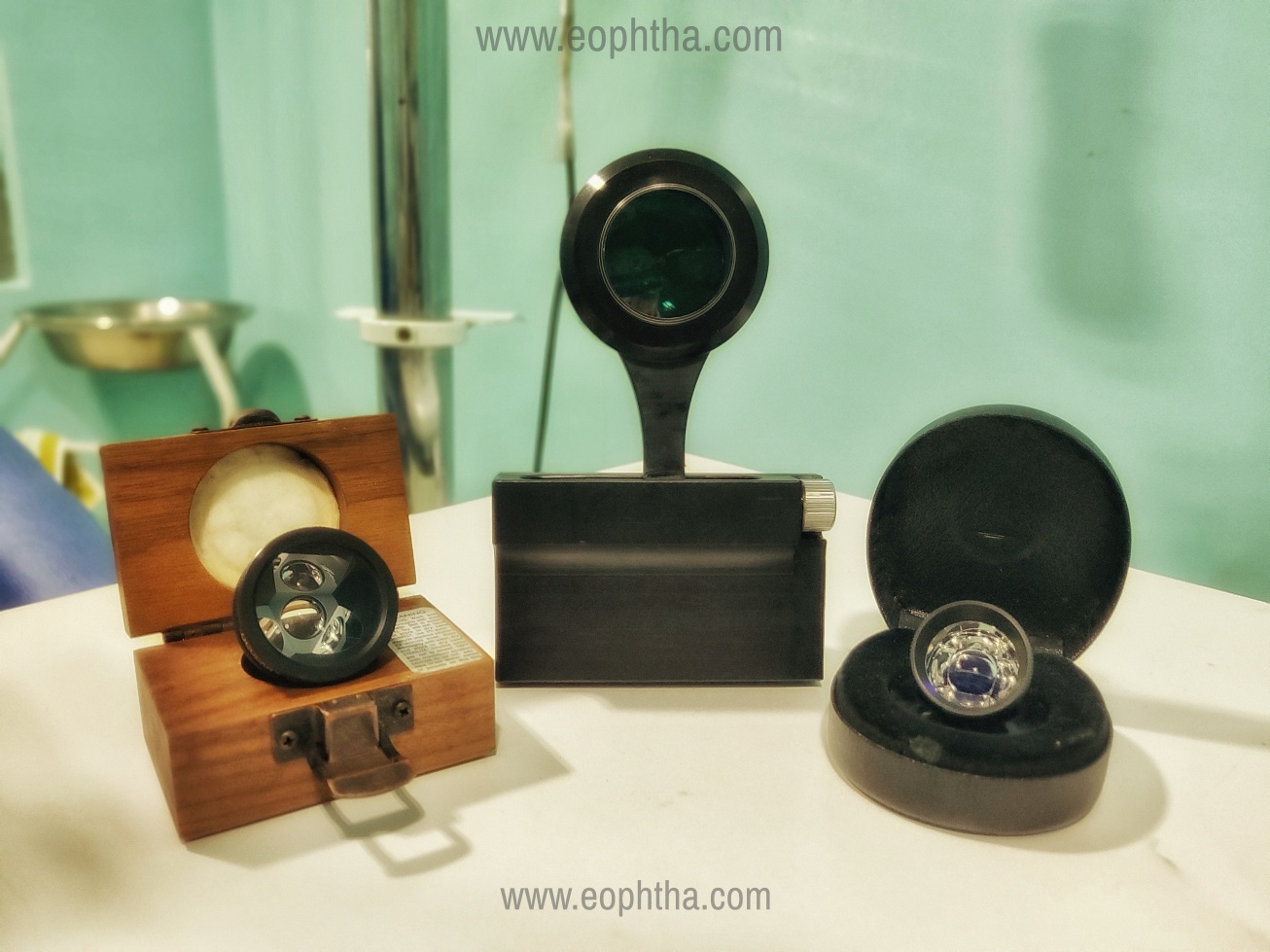
Smartphone imaging has caught the fancy of ophthalmic innovators recently. While multiple do-it-yourself (DIY) instruction videos are available on the internet for imaging ocular structures, many established players such as Keeler and Volk have forayed in the avenue. Even exclusive smartphone imaging companies are now being established to provide solutions for ocular imaging. Various DIY smartphone gonio-imaging projects have been described before[7] we published our innovative technique, later christened as Direct Imaging of Goniomirror by Smartphone (DIGS).[8] The novelty of DIGS was the slit-lamp independence, which was never described before. That made it deployable in various settings including outreach camps and patients in the supine position.
To the next level:
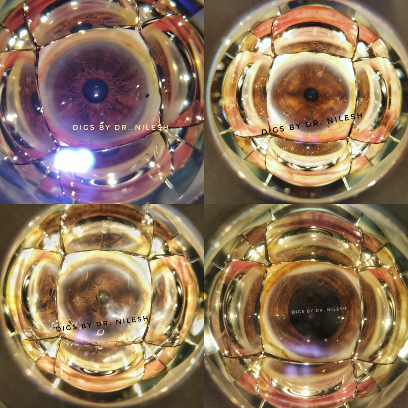
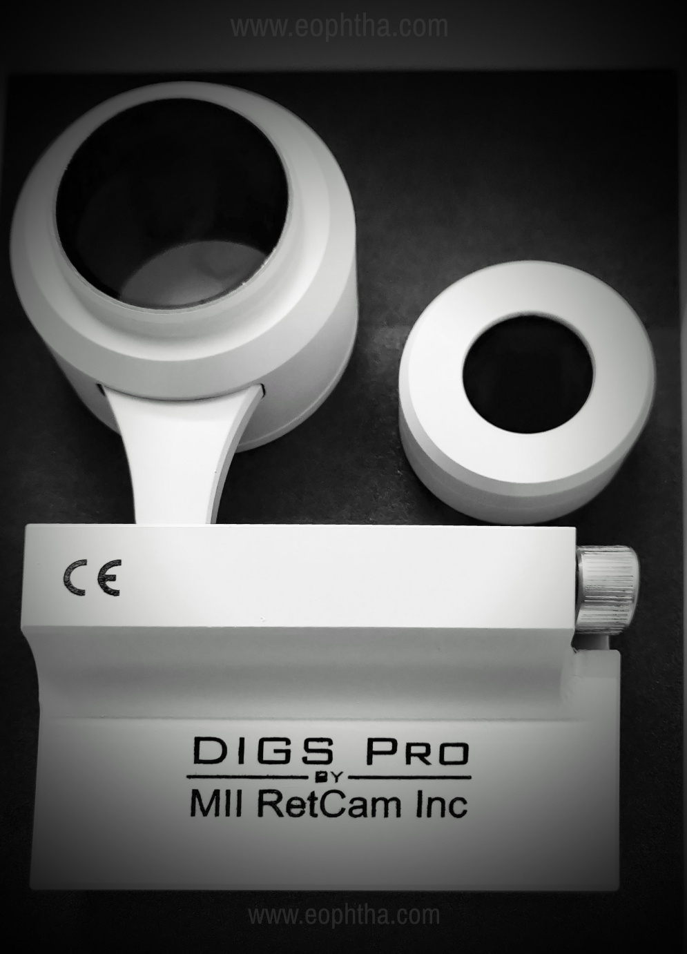
The DIY project though was lauded by the scientific community and peers, was not being used as extensively as we envisioned. The “jugaad” then graduated to an “innovative product” with the advice of my mentor Dr. Ashish Sharma, innovator of MII Ret Cam. He insisted to create a standardized product that can be easy enough to be used by ophthalmologists as well as assistants, optometrists, and technicians. The research and development of the product took almost 1 year to finalize a product that has a universal smartphone adapter along with a goniolens holder. The goniolens holder can house all the major types of 2-mirror, 3-mirror, 4-mirror, and 6-mirror goniolens. The advantage of the 4-mirror and 6-mirror lens is to provide a single picture panoramic view of the angle of the anterior chamber, providing a rapid screening method for the anatomy.[Figure 1A-D] The device is now around 1 year into the patent process and is the World’s first and to date the only smartphone-based gonio-imaging device, we like to call DIGS Pro.[Figure 2A-B]
The device has been robustly validated by correlating its finding to the slit-lamp gonioscopy findings. The inter-observer correlation was also significant in the validation study. The device has been applauded by the community and has been awarded the top prize in the “Think Under The Apple Tree” session of AIOC 2020 at Gurugram, India. The presentation of the validation study was adjudged the best presentation at “All India Talent Search Program-2020” by AIOS Scientific Committee in November 2020. It was also awarded the best innovation in the postgraduate session of “Yen-novation 2020” organised by Yennapoya University, Mangalore.
The device has found utility in patient education. The patients when shown their closed-angle with the device, agreed to the prophylactic peripheral LASER iridotomy when compared to the verbal counselling by the ophthalmologists, as found out in a study conducted recently.
The device though is limited by the smartphone camera capabilities. The gonio-anatomy contains very minute structures and thus require very high-resolution images. The current smartphone camera sensors do not provide big enough pixels to diagnose subtle findings like neovascularization of the angle or fine Sampaolesi’s line. DIGS Pro thus currently is a screening device to classify open and closed angle rather than the diagnostic device it holds the potential to be. With the in-built internet capabilities, it can be used for teleconsultation, and with high processing capabilities, the machine learning algorithm can automatically classify the images to angle-closure suspects or angle’s structural abnormalities.
References:
1. Alward WLM. A History of Gonioscopy. Optom Vis Sci 2011;88(1):29–35. 2. AEH Pondicherry. Gonioscopy: Still State of the Art After 100 Years? - Dr. Wallace Alward [Internet]. 2020 [cited 2021 Mar 7]. Available from: https://www.youtube.com/watch?v=zwNQLOfBGn8 3. Hertzog LH, Albrecht KG, LaBree L, Lee PP. Glaucoma Care and Conformance with Preferred Practice Patterns. Ophthalmology 1996;103(7):1009–13. 4. Tham Y-C, Li X, Wong TY, Quigley HA, Aung T, Cheng C-Y. Global Prevalence of Glaucoma and Projections of Glaucoma Burden through 2040. Ophthalmology 2014;121(11):2081–90. 5. Garudadri C, Senthil S, Khanna RC, Sannapaneni K, Rao HBL. Prevalence and Risk Factors for Primary Glaucomas in Adult Urban and Rural Populations in the Andhra Pradesh Eye Disease Study. Ophthalmology 2010;117(7):1352–9. 6. Faschinger C, Hommer A. Gonioscopy. Berlin: Springer; 2012. 7. Akkara J, Kuriakose A. How-to guide for smartphone slit-lamp imaging. Kerala J Ophthalmol 2019;31(1):64. 8. Kumar N, Francesco B, Sharma A. Smartphone-based Gonio-Imaging: A Novel Addition to Glaucoma Screening Tools. J Glaucoma 2019;28(9):e149–50.