
Retinopathy of Prematurity (ROP) is a proliferative disease of the retina which affects developing retinal vessels of prematurely born infants. The screening process should be carried out within 1 month of delivery of the infant. Delaying this or missing out on screening could result in the progression of abnormal neovascularization, which can lead to the development of tractional retinal detachment in the absence of treatment and this could result in irreversible blindness. There are various aspects one needs to take care of while carrying out ROP screening. Here are a few practical tips for carrying out an effective screening and follow-up in premature infants for ROP. This might be useful for paediatricians / hospital staff as well, to understand the importance of screening and ways to aid in carrying out a successful screening procedure.
1. Infant to be screened: Whom to screen?
Different countries have different screening guidelines. Ours is a developing nation with even bigger infants reported to have ROP.[1] The National Neonatology Forum of India has laid down screening guidelines.[2]
According to guidelines, any infant born:
- At < 34 weeks and/or who weighs < 1750 grams needs to be screened for ROP.
- Apart from this, any infant with 34-36 weeks and 1750-2000 grams can also be screened based on systemic risk factors for ROP.
2. Timing of screening: When to screen?
Ophthalmologists and Neonatologists both are equally responsible for the timely screening of the infant. Initial awareness among the parents and information regarding the screening has to be taken care of by the neonatologist and hospital staff. Any and every premature infant admitted to a pediatric ward or Neonatal Intensive Care Unit (NICU), falling under screening guidelines [3]
- Needs to be screened ideally at 4 weeks of delivery.
- If an infant is born < 28 weeks or is < 1200 grams, the screening procedure needs to be carried out at 2-3 weeks as these infants are at higher risk of developing a severe form of ROP, APROP, which might progress faster and earlier than 4 weeks.
3. Place of screening: Where to screen?
The ideal place for screening is in NICU, in the presence of neonatologist and assisting nurse. A monitor should be available to keep a check on vitals while screening the infant. Apart from this, infants can also be screened at an ophthalmic set up by a trained ROP specialist, if the infant is being discharged before the screening date. Care has to be taken that there is a warm and clean environment.
4. Procedure of screening: How to screen?
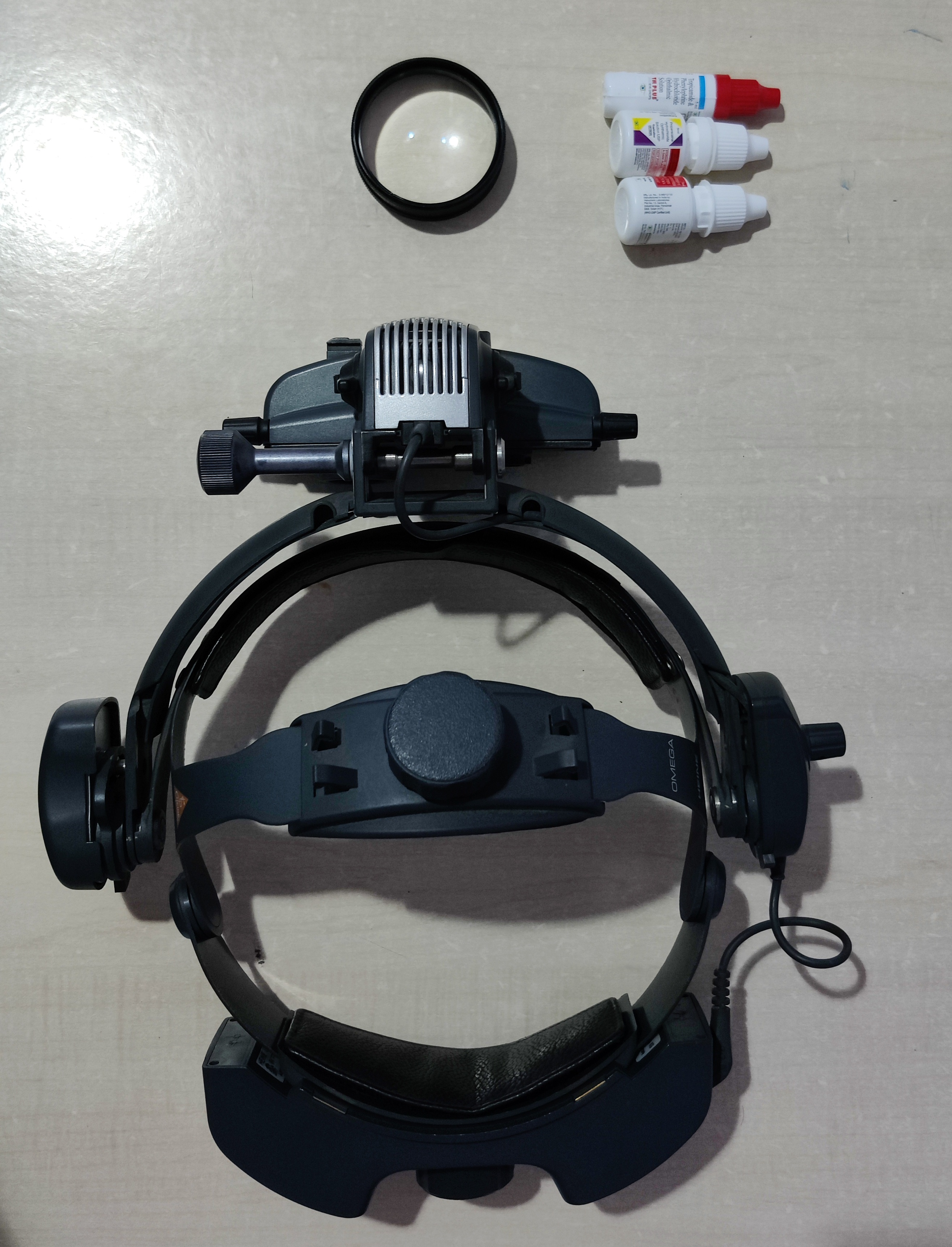
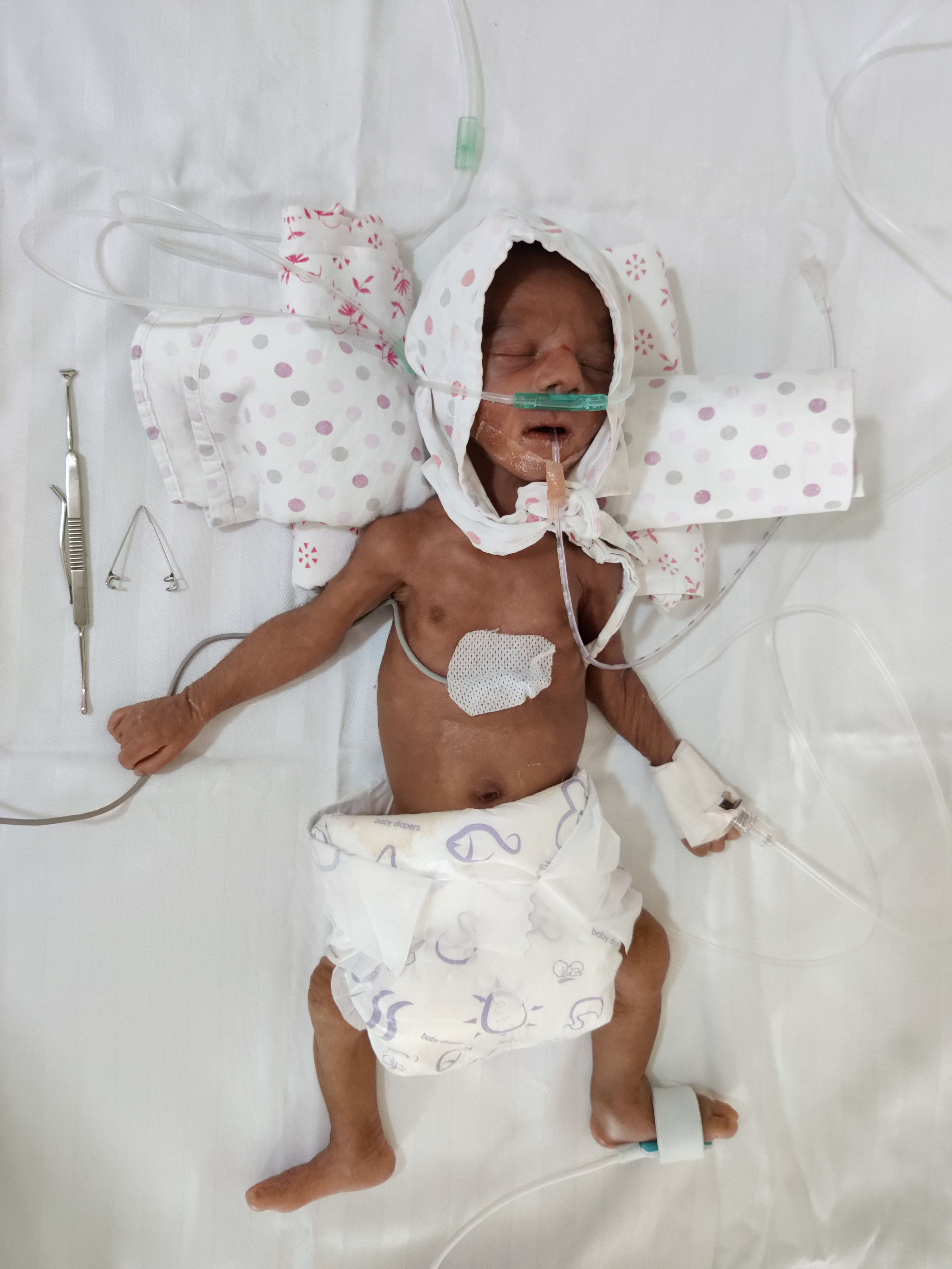
It’s better to not feed the infant an hour before screening or procedure; to avoid undue vomiting and chances of aspiration while screening or treating. Hence, clear instruction to the nursing staff should be given so that parents can be prepared well in advance. One needs to instil a Mydriatic eye drop 3 times at an interval of 10 minutes, half an hour before screening. Tropicamide 0.4% and Phenylephrine 2.5% (half the adult formulation) can be prepared by mixing an equal volume of a lubricating eye drop in the adult formulation. Now a days few pharmaceutical companies have come up with ready formulations for ROP screening, which are readily available and can be made use of directly without intermixing. For screening, the ROP specialist needs to carry an indirect ophthalmoscope, 20 D lens, Alfonso speculum, and scleral depressor. Among the other topical medications, we need an anesthetic agent (Proparacaine 0.5%), a lubricating eye drop (carboxymethyl cellulose 0.5% or 1%), and an antibiotic agent (tobramycin 0.3%) as well. [Figure 1,2] Alfonso speculum needs to be placed and with the help of a scleral depressor both eyes need to be screened, one after the other, with a 360-degree retinal evaluation.
5. Evaluation of eyes: What to screen?
General evaluation of ocular adnexa, anterior segment assessment of cornea, and anterior chamber need to be done. Comment has to be made on the status of iris neovascularisation (if any), pupillary dilatation (good/poor), and lens status (presence of TVL or lenticular opacity). This needs to be followed by a comment on media clarity. Comment on Optic Nerve Head, vasculature arising from the ONH, extent of vascularisation, presence or absence of plus disease, has to be made. Further comment has to be made on the presence or absence of any stage of ROP, with clock hours of involvement. Identification of neovascularisation along with vascular loops and avascular retina beyond that is important to look for its most aggressive variant, APROP. Staging of disease has to be done based on ICROP classification.[2] On follow-up, one needs to assess the extent of vascularisation, the status of plus disease, recurrence of retinopathy. If the laser has been done, then one must look for any ‘skip areas’ which might have to be subsequently lasered.
Follow up guidelines
|
Zones of retinal findings |
Stage of retinal findings |
Follow up interval |
|
Zone 1 |
Immature Vascularisation |
1-2 weeks |
|
Stage 1 or 2 |
1 week or less |
|
|
Regressing ROP |
1-2 weeks |
|
|
Zone 2 |
Immature Vascularisation |
2-3 weeks |
|
Stage 1 |
2 weeks |
|
|
Stage 2 |
1-2 weeks |
|
|
Stage 3 |
1 week or less |
|
|
Regressing ROP |
1-2 weeks |
|
|
Zone 3 |
Stage 1 or 2 |
2-3 weeks |
|
Regressing ROP |
2-3 weeks |
Treatment Guidelines (as per ETROP guidelines)[4]
|
Zone 1 |
No PLUS |
Stage 1 |
Follow |
|
Stage 2 |
Follow |
||
|
Stage 3 |
Treat |
||
|
PLUS |
Stage 1 |
Treat |
|
|
Stage 2 |
Treat |
||
|
Stage 3 |
Treat |
||
|
Zone 2 |
No PLUS |
Stage 1 |
Follow |
|
Stage 2 |
Follow |
||
|
Stage 3 |
Follow |
||
|
PLUS |
Stage 1 |
Follow |
|
|
Stage 2 |
Treat |
||
|
Stage 3 |
Treat |
6. Completion of follow-up: When to terminate screening? [5]
- Complete retinal vascularization; usually occurs at about 40-42 weeks of postmenstrual age.
- Infants with a post-menstrual age of 45 weeks with no evidence of ROP.
- Spontaneous Regression of ROP noted.
7. Documentation: What all to write?
Important is documentation of each finding clearly. This is important for 2 specific reasons. First, it helps you in evaluating fundus and corroborating your findings more effectively on follow-up. You can plan your management well if you have well-documented findings, which include detailed demographics along with birth history and systemic risk factors, a note of clinical findings including retinal diagrams whenever possible. You can track the progression of the disease with all these details and plan further course of treatment and follow-up. A signed copy of the findings and follow-up schedule should be handed over to the parents and another copy to be kept in ROP specialists' records. Second, it helps in being safe medico legally. Any lapse in the follow-up or scheduled treatment plan from the parents' side can lead to progression of disease in absence of the infant undergoing advised treatment, this in turn; can lead to progression of the disease and irreversible blindness. Parents often tend to blame the consulting ROP specialist for this and may file a lawsuit against the treating doctor. In a recently published perspective by Vinekar et al.[6], all the cases that have been filed against the doctors in treating infants with ROP in India, has been very well highlighted. This can very well be prevented if well documented papers are presented in the court of law. Final report being handed over to the parents should have details about diagnosis, follow-up date, and place of a visit, along with possible management plan and prognosis. Apart from this, imaging of the fundus (if the facility is available) using a wide-field / pediatric retinal imaging camera needs to be done, to explain and counsel the parents in a better way and also for better documentation.
8. Consent form: Do we need permission to see an infant?
Yes, very much needed. Consent forms should ideally be signed at every step, by the parents. This includes consenting for first-time screening, evaluating on follow-up, along with details of examination, all in a single consent form. Consent form for treatment plan let it be injection; laser or surgical intervention should be separately taken. In case you advise treatment for any infant and parents deny the treatment, for whatever reason, then parents should be made to sign consent for denial of treatment. All these consent forms are important and details of every procedure being performed should be mentioned in detail and signed both by parents and treating ROP specialists. In case if the parents are unavailable during the screening then the treating neonatologist should be informed and consent should be signed by them as well.
9. Handle with care:
Every infant you examine needs to be handled carefully. One needs to take care of last feeding time if the infant is properly wrapped with the cloth around or not; to keep it warm. Ask the assistant to hold the infant tightly while you screen, as infants, might roll towards the edge of the cradle or table if not kept in check. A topical anesthetic agent should be instilled before placing the speculum. As the speculum is placed for a longer time, one needs to maintain corneal hydration with a lubricant. Any amount of excess drops needs to be wiped off from the infant’s face. Adverse effects of ROP screening should also be kept in mind. Systemic adverse effects include Tachycardia or bradycardia, fall in oxygen saturation, apnoea, vomiting and aspiration, feeding difficulties. Ocular adversities include chemosis, subconjunctival hemorrhage, and conjunctival tear. NICU nursing staff should be asked to keep a check on the infants, post-screening for any systemic adverse events.
10. Counselling and reassurance: Can my assistant do that for me?
Well, I don't think so. These are newly born premature infants. Already the parents may be in trouble as the infant must have some associated systemic issues for which the infant is either on supplemental oxygen for maintaining saturation or have had a blood transfusion for low hemoglobin or be on systemic antibiotics for sepsis. Now to add to that we screen them for ROP and the eye is a sensitive part; parents are all the more apprehensive. So you need to counsel and explain to every parent separately, explain to them what ROP is, what is the importance of screening is, and follow up as well. They should be made mentally prepared if you suspect the need for treatment on follow-up regarding the available treatment options and what is the most suitable option for their infant. Each and every query of the parents needs to be answered peacefully without rushing into assessing the next infant or patient.
References:
- Sanghi G, Dogra MR, Katoch D, et al. Aggressive posterior retinopathy of prematurity in infants ≥ 1500 g birth weight. Indian J Ophthalmol. 2014;62:254-7.
- Project operational guidelines. Prevention of Blindness from Retinopathy of Prematurity in Neonatal Care Units. Available from: https://www.ontop-in.org/ontop-pen/Week-12-13/ROP%20NNF%20Guidelines%20.pdf [Last accessed on 25th September 2021]
- International Committee for the Classification of Retinopathy of Prematurity. The International Classification of Retinopathy of Prematurity revisited. Arch Ophthalmol. 2005;123:991-9.
- Early Treatment for Retinopathy of Prematurity Cooperative Group. Results of the early treatment for retinopathy of prematurity randomized trial. Arch Ophthalmol 2003;123:991-9.
- Section on Ophthalmology American Academy of Pediatrics; American Academy of Ophthalmology; American Association for Pediatric Ophthalmology and Strabismus. Screening examination of premature infants for retinopathy of prematurity [published correction appears in Pediatrics. 2006;118:1324]. Pediatrics. 2006;117:572-6.
- Vinekar A, Gangwe A, Agarwal S, Kulkarni S, Azad R. Improving Retinopathy of Prematurity Care: A Medico-Legal Perspective. Asia Pac J Ophthalmol (Phila). 2021;10:437-41.