
The word “serpiginous” is an adjective which means “with a wavy or indented margin”. The serpiginous choroiditis shows the similar wavy or amoeboid like lesions in choroid as a result of inflammation of unknown aetiology. The serpiginous choroiditis may be defined as a chronic, progressive, usually bilateral, recurrent in?ammatory condition which affects the choroid, choriocapillaries and retinal pigment epithelium. This clinical entity was first reported by Junius in 1932 who termed it as “peripapillary retinochoroiditis”. Thereafter this clinical entity has passed through various nomenclatures by various authors (Table 1). The term “serpiginous choroidopathy” was coined by Gass in 1987.
|
Table 1: Different nomenclatures of Serpiginous choroiditis by various authors
|
Serpiginous choroiditis is a rare cause of posterior uveitis, usually less than 5% in most of the studies from the world1.However, it has been reported in various studies 2, 3, 4, 5 that the incidence of serpiginous choroiditis is higher in India.(Table 2) The disease affects healthy, young to middle aged adult with higher male predominance. Though there is no familial predisposition, in one study the clinical entity was found to be associated with HLA B7 6.
|
Table 2: Reported incidence rate of serpiginous choroiditis in various Indian studies2,3,4,5 |
Aetiopathogenesis:
The etiology of serpiginous choroiditis is unknown. Association of various infective agents has been implicated in aetiopathogenesis of this clinical entity. A role of possible viral aetiology has been suggested by Gasset al7who reported a case of serpiginous choroiditis following Herpes zoster. Priyaet al8 reported that two-thirds of aqueous humor samples from patients with serpiginous choroiditis in their study were positive for varicella zoster virus (VZV) or herpes simplex virus (HSV) using the polymerase chain reaction. However the serological evidences in most of the study do not suggest such association and also treatment with antiviral drugs were not effective in majority of the studies. A subtype of serpiginous choroiditis, especially in India, may have active tuberculous choroiditis or an autoimmune choroiditis related to tuberculosis. Guptaet al9 reported 7 cases of ocular tuberculosis who presented with serpiginous choroiditis and showed considerable improvement in terms of visual acuity and clinically, when treated with antitubercular drugs. Laatifkainen and Erkkila 10 reported 9 patients with serpiginous choroiditis and all of them had positive tuberculin skin tests. With advent of newer diagnostic tests like interferon (IFN- γ) release assays- QuantiFERON TB gold tests, the diagnosis of tubercular infection has become easier and more accurate. QuantiFERON is an indirect test forM. tuberculosisinfection. The test has a higher speci?city than the tuberculin skin test, as it is unaffected by prior BCG vaccination, and similar or even better sensitivity. With the help of this test, Friederikeet al11 reported that 11 of 21 serpiginous choroiditis patients (52%) were tested positive in their study, indicating a tuberculous etiology in this uveitis entity. There are also reports of an immune-mediated mechanism attributable to HLA-B7 and retinal S antigen associations 6, 12. An elevation of factor VIII-von Willebrand antigen has been found in a small series of patients.13
Histopathogical Features7, 14, 30:
The characteristic finding of the lesions in serpiginous choroiditis are atrophy of the choriocapillaries, retinal pigment epithelium and photoreceptors. Among them most affected structures are choriocapillaries, which appear acellular. The larger choroidal vessels are generally spared. Lymphocytic infiltrates are seen near the margins of the lesions. Among the areas of extensive retinal pigment epithelium atrophy, there are few areas of RPE hypertrophy which is clinically seen as pigment clumping in healed lesions.
Clinical Pictures:
Serpiginous choroiditis is a bilateral condition with asymmetric involvement of the eyes. The patient typically presents with unilateral decrease in vision, photopsias, metamorphopsia, and visual ?eld loss. On examination, the anterior segment is usually normal. If present, the vitritis is mild and most often in the form of pigmented cells in anterior vitreous. Serpiginous choroiditis involves the peripapillary region and macula. Though many authors categorise this clinical entity as a white dot syndrome, the lesions in serpiginous choroiditis are not typically multifocal and most often they don’t resemble the classical white dots in fundus 30. Recurrences are common and can occur weeks to years after the initial event. Depending on the extent of lesions, serpiginous choroiditis can be divided in to the following types:
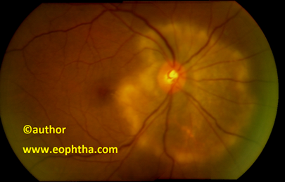
Classic or Peripapillary GeographicSerpiginous choroiditis:
Classic or Peripapillary Geographic variety accounts for 80% cases of serpiginous choroiditis. The lesion begins with ill-defined patches of grayish or creamy yellow subretinal infiltrates which starts at the peripapillary area and progresses towards the periphery like a serpentine in a centrifugal manner.(FIGURE 1,2) The overlying retina is secondarily involved and becomes oedematous .Though rare, sometimes serous exudative detachment can occur 15. The active lesions generally resolve within 6-8 weeks with or without treatment, ultimately leaving behind areas of choriocapillaries and retinal pigment epithelium atrophy. (FIGURE 3) Typically multiple lesions in different stages of resolutions are seen in fundus (Table-3). Many a time, the disease remains asymptomatic until the fovea is affected. It has been seen that about two third of patients with serpiginous choroiditis may present with scars or healed lesions at the time of initial presentation 16.
Classic or Peripapillary Geographic Serpiginous choroiditis
Macular Serpiginous choroiditis:
Macular variety, accounts for 5.9% cases of total serpiginous choroiditis cases. 7 The lesion begins in the macular area and is characterized by worse visual prognosis due to foveal involvement and higher risk of secondary CNVM. This variety of serpiginous choroiditis is often remains under-diagnosed or misdiagnosed as other macular conditions like age related macular degeneration ,toxoplasmosis etc.
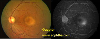
Colour and red free fundus photograph of Macular Serpiginous choroiditis
Ampiginous Choroiditis:
This is a rare variety of serpiginous choroiditis where the lesions generally occur in periphery in a multifocal pattern. Ampiginous choroiditis was first reported by Lyness and Bird in 1984, who described a recurrent form of acute posterior multifocal placoid pigment epitheliopathy (APMPPE) that resembles serpiginous choroiditis in its bilateral nature, fluorescein angiographic features, resultant pigmentary disturbances and the recurrent clinical course18. The term “Ampiginous Choroiditis” was coined by Nussenblattet al. Occasionally the acute posterior multifocal placoid pigment epitheliopathy may show lesions like serpiginous choroiditis (FIGURE 3) which indicates that two disease may represent different parts of the clinical spectrum of same condition.
Compared to the classic variety of serpiginous choroiditis, there is no significant difference in anterior segment inflammation, vitritis in Ampiginous Choroiditis. However the central foveal involvement is less in Ampiginous Choroiditis.
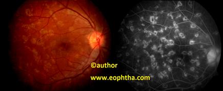
Ampiginous Choroiditis
|
Table 3: Active and Healed Lesions of Serpiginous choroiditis
|
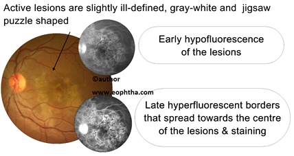
Characteristic active lesion in serpiginous choroiditis
Characteristic healed lesion in serpiginous choroiditis
Ancilliary Tests:
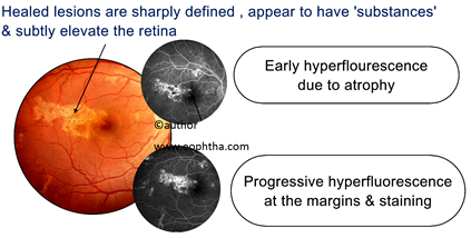
Fundus fluorescein angiography:FFA shows early hyper?uorescence of the active lesions. The late phase of the study demonstrates hyper?uorescence of the border of the active lesion that may extend centrally (FIGURE 4). Inactive lesions show early hyperflourescence and progressive hyperfluorescence with late staining of the sclera and scar tissue (FIGURE 5).
Indocyanine Green angiography:ICG is often more sensitive than FFA in determining extent and appearance of new subclinical lesions. Some authors reported that the lesions which were not apparent on FFA can be detected with the help of ICG 19, 20.Also ICG shows larger area of hypofluorescence in active lesions than seen clinically or on FFA.ICG in serpiginous choroiditis shows hypofluorescent areas beginning from early to late phase indicating non perfusion of the choriocapillaries and often areas of delayed filling, indicating late perfusion of the choriocapillaries. Also choroidal neovascular membrane may be better differentiated in areas of scar since there is minimal to no staining of scar unlike FFA.
Visual fields & Electrophysiological Studies:Visual field shows absolute or relative scotomas corresponding to the lesions .Electrophysiologic studies are usually normal.
Differential Diagnosis:
Though serpiginous choroiditis is easy to diagnose from its characteristic lesions and pattern of involvement, few conditions which affect choriocapillaries like acute posterior multifocal placoid pigment epitheliopathy (APMPPE), ocular tuberculosis, toxoplasmosis and multifocal choroiditis may need to be differentiated from serpiginous choroiditis for similar clinical and angiographic pictures as their management differs significantly.
Acute posterior multifocal placoid pigment epitheliopathy :
Like Serpiginous choroiditis, acute posterior multifocal placoid pigment epitheliopathy(APMPPE) is a bilateral condition which affects the young adults. The pattern of involvement in APMPPE is more symmetrical than serpiginous choroiditis. An antecedent viral illness has been reported in one third cases of APMPPE. The characteristic lesions in APMPPE are multiple, flat, yellow white or cream coloured plaques which are usually less than one disc diameter and of varying size, but clearly defined. The lesions typically begin in posterior pole and progresses to periphery but the lesions do not extend beyond the equator. In later stages, the lesions are replaced by varying degrees of RPE atrophy and hyperpigmentations. Both the lesions of APMPPE and serpiginous choroiditis bear the same fundus fluorescein angiographic features - early hypofluorescence and late hyperfluorescence. However, visual prognosis in APMPPE is excellent with spontaneous recovery of visual acuity to 6/12 or better within 3 to 6 weeks and recurrences are rare.
Toxoplasmosis :
Sometimes it is very difficult to differentiate a toxoplasma chorioretinal scar from healed lesions of serpiginous choroiditis because the new active lesions in both the conditions occur near the margin of old healed lesions and both the condition share same angiographic characteristics. However unlike serpiginous choroiditis, the ocular toxoplasmosis is associated with significant amount of vitritis and the patients have increased toxoplasma antibody titers.
Ocular Tuberculosis:
Both the conditions affect choroid resulting in similar scarred lesions. Many a time the serpiginous choroiditis like condition may arise in patients with tuberculosis. Guptaet al9 reported 11 eyes in 7 patients with the diagnosis of choroidal tuberculosis who presented with serpiginous choroiditis like lesions with strongly positive tuberculin skin tests and positive findings in chest radiographs. Four out of 11 eyes presented with choroidal lesions typical of serpiginous choroiditis, another 4 out of 11 eyes presented with multifocal lesions like ampiginous choroiditis and the remaining 3 eyes presented as mixture of both. These reflect the difficulty in differentiating these two conditions. However, patients with ocular tuberculosis frequently present with vitritis, constitutional symptoms, such as loss of weight, appetite, and fever, other systemic involvement, a positive tuberculin skin test and positive QuantiFERON TB gold test.
Multifocal choroiditis and Panuveitis:
The lesions in Multifocal choroiditis and Panuveitis are smaller in size, which in later stages often leaves punched out scars and they are distributed throughout the fundus, predominantly in posterior pole. Though both Multifocal choroiditis and serpiginous choroiditis shares similar angiographic characteristics, they can be differentiated by the presence of anterior uveitis, significant vitritis and smaller size of lesions in former.
Treatment:
Depending on the various proposed theory of aetiopathogenesis, several treatments have been tried for serpiginous choroiditis, but the low incidence of the clinical entity has precluded a randomized treatment trial to date.
Systemic corticosteroids are found to be effective in controlling the active lesions and shortening the duration of active diseases 21 but their role in prevention of recurrence is doubtful. Serpiginous choroiditis has been reported to recur while tapering and discontinuation of systemic corticosteroids 22 thereby emphasizing the role of long term corticosteroids therapy. However in cases of fovea-threatening lesions, aggressive rapid control of the in?ammation is needed as it has been reported that the response of serpiginous choroiditis to oral steroids occurs after two weeks of treatment 23. So, high-dose intravenous steroid therapy (1 g intravenous methylprednisolone daily for three days) is recommended by many authors for macula-threatening cases of serpiginous choroiditis23.There are also reports of intravitreal Triamcinolone Acetate (IVTA) therapy in serpiginous choroidopathy26. IVTA was also used in cases where systemic corticosteroids were contraindicated 24 and in a case of secondary choroidal neovascular membrane25.It has been observed that though IVTA injection brings in the required concentration of the drug without systemic side effects to the desired tissue level and likely to be effective in the treatment of acute lesions, but it is not helpful in preventing recurrence of the disease 26.
The spectrum of alternative therapies to systemic corticosteroid treatment ranges from immunosuppression with cyclosporine alone or as part of a regimen with immunosuppressives and most of the study using these agents showed mixed result. Christmaset al22 reported 4 out of 6 patients with serpiginous choroiditis treated with 2–40 months of immunosuppressive drugs, such as cyclosporine A, azathioprine, or mycophenolate mofetil, successfully discontinued their therapy without recurrences. On the other hand, Akpeket al27 reported that 2 out of 4 patients with serpiginous choroiditis in their study, who were treated with cyclosporine alone or combined with azathioprine experienced a recurrence while on therapy.
A triple-agent immunosuppressive regimen using cyclosporine (5mg/kg/day initially) in combination with azathioprine (1.5mg/kg/day) and prednisone (1mg/kg/day) was first reported by Hooper and Kaplan in 1991 28. Treatment was tapered 8 weeks after initiation and discontinued after 6 months, when no recurrence was encountered. However, as the medications were weaned, recurrence of the inflammation developed in two patients. Munteauet al17 also reported satisfying results with this triple-agent therapy. Although prompt control of the inflammation on initiation of treatment was achieved, recurrence of inflammation after discontinuation of treatment has been reported in some studies. Also, Biswaset al29 reported that there was no significant change in the rate of regression of lesions in their study when this regimen was compared to the treatment with azathioprine or corticosteroids alone.
There are also reports of use of Interferon Alpha used in management of Serpiginous choroiditis. Sobaciet al31 reported of successfully treating 8 eyes of 5 patients with IFN alpha-2a, but they could not explain the mechanisms by which IFN affects the course of Serpiginous choroiditis.
|
Treatment of Serpiginous choroiditis: Key Topics
|
Prompt diagnosis and rapid initiation of treatment of active lesions with immunosuppression and maintenance of appropriate immunosuppression for at least 6 months is essential for initial management and prevention of recurrences in Serpiginous choroiditis. A prospective randomized clinical trial may help us to ?nd the most effective modality of treatment in patients with serpiginous choroiditis.
REFERENCES
-
Chang JH, Wakefield D: Uveitis: a global perspective. Ocul Immunol Inflamm 10:263–79, 2002
-
Biswas J, Narain S, Das D, et al: Pattern of uveitis in a referral uveitis clinic in India. Int Ophthalmol 20: 223–8, 1996-97
-
Rathinam SR, Namperumalsamy P: Global variation and pattern changes in epidemiology of uveitis Indian Journal of Ophthalmology, Volume 55, Issue 3,2007
-
Singh R, Gupta V, Gupta A: Pattern of Uveitis in a Referral Eye Clinic in North India. Indian Journal of Ophthalmology, Volume 52, Issue 2, 2004
-
Das D, Bhattacharjee H, Bhattacharyya P: Pattern of uveitis in North East India: A tertiary eye care center study, Indian Journal of Ophthalmology, Volume 57, Issue 2, 2009
-
Erkkila H, Laatikainen L, Jokinen E: Immunological studies on serpiginous choroiditis. Graefes Arch Clin Exp Ophthalmol 219:131–4, 1982
-
Gass JDM: Stereoscopic atlas of macular diseases: diagnosis and treatment, vol 1, St Louis, Mosby, ed 3, pp 136–144, 1987
-
Priya K, Madhavan HN, Reiser BJ et al: Association of herpes viruses in the aqueous humor of patients with serpiginous choroiditis: a polymerase chain reaction-based study. Ocul Immunol Inflamm 10:253–61, 2002
-
Gupta V, Gupta A, Arora S, etal: Presumed tubercular serpiginous like choroiditis: clinical presentations and management. Ophthalmology 110:1744–9, 2003
-
Laatikainen L, Erkkila ¨ H: Serpiginous choroiditis. Br J Ophthalmol 58:777–83, 1974
-
Friederike M, Matthias D. B, Ute W, Regina M, Alexander D, and Stefan Z: QuantiFERON TB-Gold—A New Test Strengthening Long-Suspected Tuberculous Involvement in Serpiginous-like Choroiditis. Am J Ophthalmol; 146:761–766. 2008
-
Broekhuyse RM, van Herck M, Pinckers AJ, et al: Immune responsiveness to retinal S-antigen and opsin in serpiginous choroiditis and other retinal diseases. Doc Ophthalmol 69:83–93, 1988
-
King DG, Grizzard WS, Sever RJ, et al: Serpiginous choroidopathy associated with elevated factor VIII-von Willebrand factor antigen. Retina 10:97–101, 1990
-
Wu JS, Lewis H, Fine SL, et al: Clinicopathologic findings in a patient with serpiginous choroiditis and treated choroidal neovascularization. Retina 9:292–301, 1989
-
Hoyng C, Tilanus M, Deutman A: Atypical central lesions in serpiginous choroiditis treated with oral prednisone. Graefes Arch Clin Exp Ophthalmol 236:154–6, 1998
-
Laatikainen L, Erkkila ¨ H: A follow-up study on serpiginous choroiditis. Acta Ophthalmol (Copenh) 59:707–18, 1981
-
Munteanu G,Munteanu M, Zolog I: Serpiginous choroiditis clinical study. Oftalmologia 52:72–80, 2001
-
Lyness AL, Bird AC: Recurrences of acute posterior multifocal placoid pigment epitheliopathy. Am J Ophthalmol 98:203–7, 1984
-
Squirrell D M ,Bhola R M ,Talbot J F . Indocyanine green angiographic findings in serpiginous choroidopathy: evidence of a widespread choriocapillaris defect of the peripapillary area and posterior pole. Eye 15:336–8, 2001
-
Van Liefferinge T, Sallet G, De Laey JJ: Indocyanine green angiography in cases of inflammatory chorioretinopathy. Bull Soc Belge Ophtalmol 257:73–81, 1995
-
Weiss H, Annesley WH, Shields JA, et al: The clinical course of serpiginous choroidopathy. Am J Ophthalmol 87:133– 42, 1979
-
Christmas NJ, Oh KT, Oh DM, et al: Long-term follow-up of patients with serpinginous choroiditis. Retina 22:550–6, 2002
-
Nikos N., Ioannis H., Simina P,Nikos G, Nikos E, Tasos K. Intravenous Pulse Methylprednisolone Therapy for Acute Treatment of Serpiginous Choroiditis Ocular Immunology & Inflammation,14:1,29 — 33,2005
-
Pathengay A. Intravitreal triamcinolone acetonide in serpiginous choroidopathy.Indian J Ophthalmol.;53:77–79, 2005
-
Navajas EV, Costa RA, Farah ME, Cardillo JA, Bonomo PP. Indocyanine green-mediated photothrombosis combined with intravitreal triamcinolone for the treatment of choroidal neovascularization in serpiginous choroiditis.Eye,17(5):563–566. 2003
-
Adgüzel, Ufuk, Sar, Ayça, Özmen, Cengiz and Öz, Özay Intravitreal Triamcinolone Acetonide Treatment for Serpiginous Choroiditis .Ocular Immunology & Inflammation,14:6,375 — 378, 2006
-
Akpek EK, Baltatzis S, Yang J, et al: Long-term immunosuppressive treatment of serpiginous choroiditis. Ocul Immunol Inflamm 9:153–67, 2001
-
Hooper PL, Kaplan HJ. Triple agent immunosuppression in serpiginous choroiditis. Ophthalmology, 98:944–951, discussion 951–952, 1991
-
Abrez, H., Biswas, Jyotirmay and Sudharshan, S. Clinical Profile, Treatment, and Visual Outcome of Serpiginous Choroiditis. Ocular Immunology & Inflammation,15:4,325 — 335,2007
-
Wee-Kiak L.Ronald R.B. Nussenblatt RB.Serpiginous Choroiditis,Survey of Ophthalmology.2005
-
Sobac, Güngör, Bayraktar, Zeki and Bayer. Interferon Alpha-2A Treatment for Serpiginous Choroiditis. Ocular Immunology & Inflammation,13:1,59 — 66. 2005