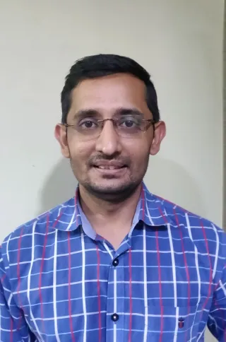
Dr. Rohit Rao

Consultant, Shri Ganesh Vinayak Eye Hospital, Raipur, Chhattisgarh.

30 articles

Dr Rohit Rao, completed MBBS from JNMC Wardha Maharashtra in the year 2010. Done MS ophthalmology from JSS MEDICAL COLLEGE Mysore Karnataka in 2015. Underwent long-term fellowship in IOL and Anterior segment under the guidance of Dr Kalpana Narendran at Aravind eye hospital Coimbatore in 2017.He underwent short term DCR fellowship from Shankara eye hospital Coimbatore under Dr Shruti Tara. Presently he is working as an Anterior Segment surgeon and oculoplasty consultant At Shri Ganesh Vinayak eye hospital, Raipur Chhattisgarh. He has quite a few National and International papers , videos and e-posters under his name. He has been very much involved in training new surgeons at Shri Ganesh Vinayak eye hospital Raipur Chhattisgarh. Dr. Charudutt Kalamkar did his MBBS and MS from AIIMS (N.DELHI). He is well versed in all aspects of clinical and surgical ophthalmology. Apart from routine phacoemulsification surgery for cataract, he is incharge of squint and glaucoma units. He also has rich experience in oculoplasty.

  1. Students Gallery 27 Dec 2024 7 min read

    Fellowship Opportunities in India for International Ophthalmologists

    Aditya Jyot Eye Hospital (ICO Sponsored Pediatric Ophthalmology, Strabismus, Cornea & External Diseases, Medical Retina, Vitreo-Retinal Diseases) Duration of Fellowship: 3 months Mode of Selection: Application via ICO website Webpage: https://www.adityajyoteyehospital.org/ophthalmic-education.html Email: researchajeh@gmail.com Contact number: Tel: 91-22-24177600 Address: Mumbai Session: Quarterly Course Fees: ICO Sponsored Arora Eye Hospital And Retina Center (ICO Sponsored Medical Retina, Cataract Surgery) Duration of Fellowship: 3 months Mode of Selection: Application via ICO website Webpage: https://eyecarehelpline.com/aroraeyehospitalandretinacenter Email: eyecarehelpline@gmail.com Contact No.: 9988610610 Address: Bangalore, India Session: Quarterly Course Fees: ICO Sponsored Dr. Solanki Eye Hospital (ICO Sponsored Cataract Surgery, Community Eye Health) Duration of Fellowship: 3 months Mode of Selection: Application via ICO website Webpage: Dr. Solanki Eye Hospital | Eye Hospital


  2. Articles 24 Jan 2023 6 min read

    Ten Points A Surgeon Should Know About the Phaco-probe

    1. Basic structure of phaco probe Phaco Probe has two attachments (For aspiration & Irrigation) and one wire coming out of back end for power supply and sensors. In the front there is the tip attached to the hand piece, which has a silicon sleeve around it with two apertures. The irrigation tubing are wider in diameter than aspiration and both tubings are colour coded Fig 1 Basic Structure of Phaco Probe 2. Fluid Inflow and Outflow pathway: The fluid flows from the irrigation tube to the handpiece and comes out via two apertures in the silicon sleeve cooling the tip. It then traverses in the AC and is aspirated back via phaco tip and comes out via aspiration tube. Irrigation is gravity based in traditional machines and Active in newer generation machines. Aspiration is always active can be via either peristaltic and venturi pump based. Fig 2 Fluid inflow and Outflow via Phaco Probe 3. Phaco Tip design: Phaco tip is made of titanium alloy. The end of the tip has a bevel, which can be of

  3. Articles 12 Jan 2022 5 min read

    Seven Tips for Sideport Construction

    Sideport might appear as a small and trivial step in SICS but it has great significance. Improperly constructed side port will give rise to a cascade of events which will complicate the further steps. You can make an ideal side port by employing the following tips. 1. Size Width: A very small side port will pose a lot of problems while instrumentation. It will force multiple attempts to enlarge the side port throughout surgery, which itself poses the risk of injury to intraocular structures. Conversely, a very big side port will lead to wound leak and instable AC intra-operatively. It may end up in needing a suture at the end. So assessment of apt width is imperative. The size of side port is largely dependent on the gauze of simcoe cannula being used. It needs a bit of experience to adequately size the side port and after doing a few cases one will be able to judge how much the blade needs to be inserted and how much extension is required. Fig 1 a. Very Big Sideport and b. Small Sidep

  4. Articles 17 Nov 2021 5 min read

    Ten Pearls for Tunnel Construction in SICS

    SICS is the most commonly performed cataract surgery by young budding surgeons. Young postgraduates are introduced to the world of ophthalmological surgery via SICS. Initially, every new surgeon struggles to make good scleral tunnel and capsulorhexis. These TOP TEN pearls will make young surgeons confident to achieve textbook tunnel. 1. Good Visualization: If you can see well, you can do well. Proper exposure of working field is a must. So, focus on to make adequate Conjunctiva and tenon’s dissection, avoid pooling of blood with judicious cautery and don’t let saline or visco to pool. Fig.1 Adequate Scleral exposure for tunnel construction 2. Superior rectus bridle suture: Mind the rectus It is probably one of the most important step in SICS. It is the first step any beginner surgeon learns, but it has not been given the due credit. It is a hidden player in tunnel construction. Furthermore, it provides the globe with sufficient traction and support to construct the tunnel seamlessly. 3

  5. Students Gallery 2 Apr 2021 4 min read

    Tips To Achieve Ideal Rhexis

    Capsulorhexis was pioneered and popularized by Howard Gimbel. Capsulorhexis is probably the most challenging to learn in cataract surgery, in other words, once a good rhexis is done half the battle is won. All the Post- Graduates and beginners struggle to do rhexis. This article has compiled small tips and tricks to do rhexis better. Ideally, rhexis should be continuous and curvilinear i.e Circular, Central & of Correct size (5.5-6 mm). Circular: for proper IOL centering and stabilization in the bag Continous: to prevent rhexis extension Correct size: for IOL centering and to ensure anterior capsule overlapping IOL optic 360 degrees. It can be done with the help of a double bent 26 G/27 G needle or rhexis forceps. Let's break the table and use the advantages of a needle Needle Utrata Forceps Advantages Cheap Disposable Universally available Customized Small diameter, lightweight Can be done through side port so less chance of AC shallowing Precise control Disadvantages Less precise con

  6. Articles 2 Apr 2021 8 min read

    A Beginners Guide to Ophthalmic Surgical Microscope

    An operating surgical microscope is the most important optical instrument in the modern era of ophthalmic surgeries. It provides the surgeon with a magnified and illuminated high-quality image of the small ophthalmic structures. Being binocular the surgical microscopes gives the additional benefit of high-quality stereoscopy. History The first German doctor to use a stereomicroscope was Dr. Hans Hinselmann (1884-1959), Prof. of Gynaecology under Dr. Otto von Franque at the University Clinic, Bonn, who adapted in 1925 a Leitz stereomicroscope with coaxial illumination to a movable floor stand. He named IT “colposcope” and used it to visualize the female genital organs particularly for cancer of the cervix. Dr. Hans Littmann (1908 – 1991), head of Med-Lab at Zeiss OptonOberkochen, West Germany (now Carl Zeiss A.G.) and his team of technicians designed in 1953 their first ophthalmic operating microscope Prerequisites for an ideal operating microscope Must have stereoscopic visualization.

  7. Interesting Reads 1 Apr 2021 2 min read

    Covid Time: Standard Operating Procedures of Various Ophthalmic Societies & Institutes

    COVID-19 pandemic has eclipsed the earth. And we need to fight it together; together with our joint efforts, we can defeat this pandemic. As ophthalmologists, we are one of the frontline commandos against it. From eOphtha, we have tried to compile various standard operating procedures and guidelines being followed by various ophthalmic and optometry practices all over the world. Kindly click the links below. Please email your recommendations and guidelines for Ophthalmologists or Optometrists during the COVID-19 pandemic to drrohit999@gmail.com Standard Operating Procedures and Guidelines All India Ophthalmological Society (AIOS): AIOS Operational Guidelines for Ophthalmic Practice during COVID 19 outbreak American Academy of Ophthalmology: Important Corona Virus Updates for ophthalmologists: Important Corona Virus Updates for Ophthalmologists American Society of Retina Specialists ASRS: Recommendations for Conducting Vitreoretinal Surgery During the COVID-19 Pandemic Canadian Society

  8. Interesting Reads 1 Apr 2021 11 min read

    Contemplating Dacryology

    Dacryology is the study of the lacrimal system of the human body. Dr. Rohit Rao and Dr. Bindu Malini from eOphtha spoke to Dr. Javed Ali, one of the pioneers in the field of dacryology. We hope this discussion would shed light on the field of dacryology and would answer some common queries pertaining to the surgical management of dacryocystitis. Dr. Javed Ali is the Head, Govindram Seksaria Institute of Dacryology, LVPEI, Hyderabad. He is also serving as the Hong-Leong Professor, University of Singapore, Singapore. and Professor, Friedrich Alexander University, Nuremberg, Germany, Adjunct Associate Professor, University of Rochester, New York, Senior Von Humboldt Scientist, FAU, Nurnberg, Germany. He completed his basic medical education and MS Ophthalmology from NTR University of Health Sciences, Hyderabad. He obtained his fellowship of Royal College of General Practitioners (FRCGP), UK in 2003 and fellowship of Royal College of Physicians and Surgeons of Glasgow (FRCS) in 2008. He al

  9. Students Gallery 1 Apr 2021 16 min read

    Instruments used in Cataract Surgery: Ready Reckoner for the Post Graduates

    Cataract Surgery is the most common surgery performed all over the world. Proper knowledge of surgical instruments used in cataract surgery is essential. This article gives you an overview and description of instruments commonly used in cataract surgery. Simcoe's irrigation- aspiration cannula: Design: Available in two different designs. The original model also called the direct Simcoe where aspiration is through the silicone tube and irrigation through the main hub. The surgeon holds the syringe attached to the silicone tubing which is used to generate suction for aspiration in his/her left hand and the irrigation line is attached to the main central hub. The other model is the Reverse Simcoe where irrigation is through the main hub. The surgeon uses a syringe attached to the main hub to generate suction for aspiration. Cannulas are available in different sizes: 21,22,23 gauge which has an aspiration port size of 0.5,0.35,0.3 mm respectively. These are 15 mm long in size with an angle

  10. Students Gallery 1 Apr 2021 9 min read

    Instruments Used in Oculoplasty

    Oculoplasty Instruments Oculoplasty deals with the aesthetic part of ophthalmology. Precision is the key in Oculoplasty as the outcomes depend on it. Good precision can be achieved with good knowledge of the instruments used in oculoplasty and when to use them. This article would give a brief idea on basic instruments used. Nettleship Punctum Dilator It is the instrument with very fine blunt tip, with a tapering end Uses Dilation of puncta during diagnostic and therapeutic probing Before syringing in small puncta, Diagnosing punctal stenosis, Canalicular repair with monoka stent, Silicone tube intubation Nettleship Punctum Dilator Castroviejo Lacrimal Dilator It is the instrument with Double-ended with fine and medium taper end. Uses Dilation of puncta during diagnostic and therapeutic probing Before syringing in small puncta, Diagnosing punctal stenosis, Canalicular repair with monoka stent, Castroviejo Lacrimal dilator Thudicum’s Nasal Speculum It is a thin instrument with flanges, o