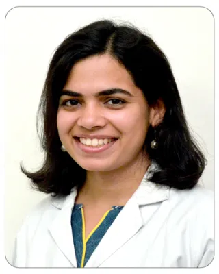
Dr. Supriya Sharma

Junior Ophthalmologist, L V Prasad Eye Institute, Banjara Hills, Hyderabad, Telangana, India, 500034, Email ID: supriyasharma@lvpei.org

2 articles

Supriya Sharma has pursued her post-graduate training in Ophthalmology followed by two-year clinical fellowship in Cornea and Anterior Segment from LV Prasad Eye Institute. Her areas of clinical interest include corneal transplantation, ocular trauma and ocular surface disease. She is keen on studying microbial keratitis and ocular surface disease, and wants to pursue these as her research interests. She has to her credit publications in peer-reviewed journals and has made several national and international presentations during her training period. Besides her clinical work, she is passionate about training post-graduate and ophthalmology fellows.

  1. Articles 8 Jul 2021 30 min read

    Anterior Segment Optical Coherence Tomography in Diseases Of The Ocular Surface And Cornea

    I. Introduction : Anterior segment optical coherence tomography (ASOCT) is a sophisticated non-contact imaging device that produces high-resolution images for detailed assessment of the structures of the ocular surface and anterior chamber in various disease conditions. Starting from the earlier generation time-domain devices first described in 1991 by Huang et al [1] to the current Fourier-domain OCT devices, the technological advancement in this device has improved the diagnostic ability tremendously. This imaging modality has evolved into a highly precise tool for clinical diagnosis and management as well as in research and understanding disease pathology. In this review, we describe the evolution of ASOCT, its technology in brief and focus on highlighting the various clinical applications with examples. While Izzat et al first introduced the use of AS-OCT for imaging the anterior segment of the eye [2], it is well known that the OCT initially evolved as a tool for retinal imaging.


  2. Articles 5 May 2021 23 min read

    Simple Limbal Epithelial Transplantation: A Review

    I. Introduction The cornea is lined by non-keratinized, stratified squamous epithelium. At the limbus, there is a gradual transition of these cells to non-keratinized, stratified columnar epithelial cells, along with mucin-secreting goblet cells, forming the conjunctival epithelium. The corneoscleral limbus lies between these two epithelia in a healthy eye. The limbus contains palisades of Vogt, the basal layer of which function as a repository for the corneal epithelial stem cells.(1, 2) These limbal epithelial stem cells (LESC’s) divide whenever the corneal epithelium is required to be replenished. As the stem cells divide, one of the daughter cells retains the primitive nature of the parent cell and functions to maintain the stem cell pool. The other daughter stem cell differentiates to acquire the physiological nature of a specific tissue, like the corneal epithelium. These cells are known as ‘transient amplifying cells’, which then divide and migrate centripetally, circumferential